
Category: IGCSE Biology posts

Thermoregulation: Grade 9 Understanding for IGCSE Biology 2.93
Homeostasis is a term that means maintaining a constant internal environment in spite of changes in the external environment. Many variables in the body are regulated by homeostasis but the two control systems specifically mentioned in your specification for iGCSE are osmoregulation (regulation of water balance) and thermoregulation (regulating of body temperature)
I have looked at osmoregulation in a previous post but in this final post for half term 2015, I will give a few details about thermoregulation.
Thermoregulation means to maintain the core body temperature at a set value. This can be energetically very costly as the animal has to respire at a much higher rate to release the heat needed to warm the body, but it has allowed mammals and birds to colonise habitats that would be inaccessible to all poikilothermic (cold-blooded) animals.
Why regulate body temperature?
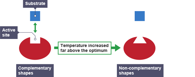
All metabolic reactions in the body are catalysed by enzymes. If the body temperature falls too low below the set value, the rate of an enzyme-controlled reaction will drop, and this would be a problem as metabolic reactions would happen too slowly. If the temperate goes much above the optimum temperature, then the enzymes that catalyse all the reactions in cells would denature. This means they will change their shape so that the “lock and key” mechanism of catalysis cannot work at all.
In any homeostatic control system there will be three components:
- Sensors (where the variable is measured)
- Integrating Centre (where the measured value is compared to a set value)
- Effectors (which can bring about a response)
In human thermoregulation, there are two sets of sensors that measure temperature. The skin contains hot and cold receptors which can respond if the skin gets too hot or cold respectively. The temperature of the blood is constantly measured by a second set of thermoreceptors which are found in the hypothalamus in the brain.
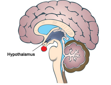
The hypothalamus also acts as the integrating centre, collecting information from a variety of sensors and then initiating an appropriate response.
The main effector organ in thermoregulation is the skin.
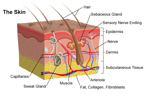
I have looked at the role of the skin in thermoregulation in an earlier post – click here to be taken to this….

Just check you understand the role of sweating, vasodilation in helping the body lose heat if it gets too hot and vasoconstriction and shivering if it gets to cold. I hope the earlier post will help!
Please add comments/feedback/questions etc using the comment feature at the bottom of this post or tweet me @Paul_Gillam.
Kidney (part II): Grade 9 Understanding of the kidney’s role in osmoregulation 2.76B, 2.78B, 2.79B
The main function of the kidneys is EXCRETION. They remove urea from the blood in a two stage process described in an earlier post, first by filtering the blood under high pressure in the glomerulus and then selectively reabsorbing the useful substances back into the blood as the filtrate passes along the nephron.
But the kidney has an equally important role in HOMEOSTASIS. It actually is the main effector organ for regulating a whole load of variables about the composition of the blood (e.g. blood pH and salt balance) but in this post I want to explain to you how the water balance of the body is regulated and the kidney’s role in this homeostatic system.
Why do you need to regulate the dilution (or water potential) of the blood?
If the blood becomes too dilute, then water will enter all the body cells by osmosis (from a dilute to a more concentrated solution). This net movement into cells would cause them to swell and eventually burst. Bad news all round…
If the blood becomes too concentrated, then water will leave the body cells by osmosis. Cells will shrivel up as they lose water into the blood and this will kill them. Bad news all round….

Remember: a hypertonic solution has a low water potential and is very concentrated. A hypotonic solution has a very high water potential and is very dilute.
The regulation of the water potential of the blood is a very important example of homeostasis in the human. It is often referred to as OSMOREGULATION.
The water potential (dilution) of the blood is measured continuously by a group of neurones in a region of the brain called the hypothalamus.
The hypothalamus is found right next to a very important hormone-secreting gland called the pituitary gland, marked as the red circular structure on the diagram above. When the hypothalamus detects that the blood’s water potential is dropping (i.e. it is getting too concentrated) this causes the posterior lobe of the pituitary gland to start secreting a hormone ADH into the bloodstream.
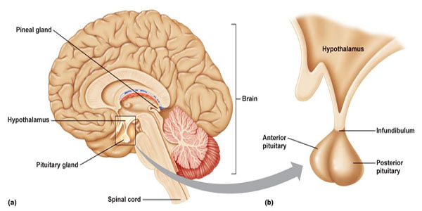
(You might remember that these brain structures appear elsewhere in the iGCSE specification. The hypothalamus also contains the temperature receptors that measure the temperature of the blood in thermoregulation; the pituitary gland plays a role in the menstrual cycle by producing FSH and LH)
Hormones such as ADH exert their effects elsewhere in the body. The main target tissue for ADH is the collecting duct walls in the kidney. ADH binds to receptors on these cells and makes the wall of the collecting duct much more permeable to water. This means as the urine passes down the collecting duct through the salty medulla of the kidney, lots of water can be reabsorbed into the blood by osmosis. This leaves a small volume of very concentrated urine and water loss is minimised.
ADH is secreted whenever the body is dehydrated. It might be because the person is losing plenty of water in sweating in which case it is vital that the kidney produces as small a volume of urine as is possible.

If you drink a litre of water, what effect will this have on the dilution of the blood: of course it makes the blood more dilute. This will be detected in the hypothalamus by osmoreceptors and they will cause the pituitary gland to stop secreting ADH into the bloodstream. If there is no ADH in the blood, the walls of the collecting duct remain totally impermeable to water. As the dilute urine passes down the collecting duct, no water can be reabsorbed into the blood by osmosis and so a large volume of dilute urine will be produced.

This is another beautiful example of negative feedback in homeostasis.
PMG tip: you can avoid getting confused in the exam about the effect of ADH if you can remember what it stands for. ADH is an acronym for anti-diuretic hormone (ADH). A diuretic is a drug that promotes urine production. They are banned drugs from WADA (World Anti-Doping Agency) since they can be taken as a masking drug to help flush out evidence of illegal drug taking. Shane Warne missed the 2003 cricket World Cup and served a ban for failing a drugs test due to diuretics in his sample.
So an anti-diuretic hormone will reduce urine production. This means it will be secreted when the body is dehydrated as the blood gets too concentrated.
Finally remember that it is not the whole nephron that is affected by ADH, just the collecting ducts and part of the distal convoluted tubules. Most water in the glomerular filtrate is absorbed in the nephron but the collecting duct has a role in “fine-tuning” the volume and dilution of urine.
This is a really important topic to master for an A* in your exam. Examiners seem to like asking questions on ADH and osmoregulation and often these questions are amongst the hardest marks to get in the exam, and so serve as a brilliant discriminator between A and A* candidates. Work hard to master this topic and with a little luck from the question-setters an A* grade is within your grasp……

Kidney (part I): Grade 9 GCSE Understanding of kidney’s role in Excretion 2.72B, 2.73B, 2.74B, 2.75B, 2.76B, 2.77B
Excretion is defined as “the removal of waste molecules that have been produced in metabolism inside cells”. So for example carbon dioxide is a waste product of respiration and is excreted in the lungs.
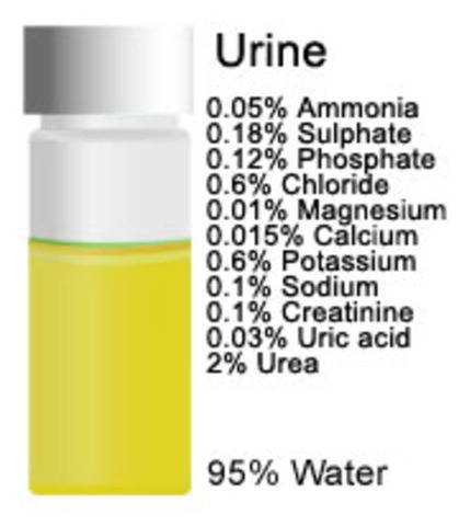
The liver too produces a waste molecule urea from the breakdown of amino acids. Amino acids and proteins cannot be stored in the body: if you eat more than you use, the excess is broken down to urea. Urea would certainly become toxic if it was allowed to accumulate in the body (patients with no kidney function will die within 3-4 days without treatment) and the organ that is adapted to excrete urea from the blood is the kidney. Kidneys excrete urea by dissolving it in water, together with a few salts to form a liquid called urine.
Don’t confuse urine, the liquid produced in the kidney that is removed from the body, with urea, the nitrogen-containing chemical made in the liver that ends up as one component of urine.
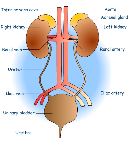
Urine is produced in the kidneys continuously day and night. It travels away from the kidney in a tube called the ureter. Each kidney has a ureter coming out of it, and the two ureters carry the urine to the bladder. The bladder is a muscular storage organ for urine. Urine drains from the bladder through a second tube called the urethra.
Make sure you check your spelling: ureter and urethra are easy to muddle and correct spelling is essential to ensure the meaning is not lost….
How is urine made in the kidney?
Well that’s the big question for this post. How does the kidney start with blood and produce a very different liquid called urine from it….. Urine is basically made of water, dissolved urea and a few salts.
Before I can explain how urine is made, I need to briefly look at the structure of a kidney.
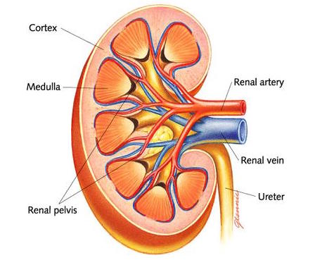
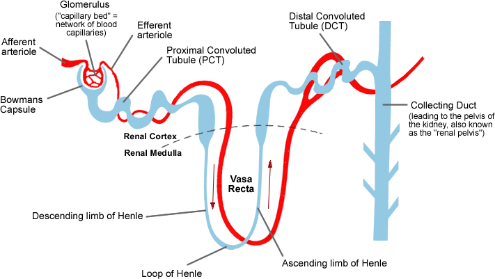
You can see the structure of the kidney on this simple diagram. There are three regions visible in a kidney: an outer cortex, an inner medulla which is often a dark red colour due to the many capillaries it contains, and a space in the centre called the renal pelvis that collects the urine to transfer it into the ureter. Blood enters the kidney through the large renal artery and deoxygenated blood containing less urea leaves the kidney in the renal vein.
But there is no way from looking at the gross structure of the kidney that you could ever work out how the Dickens it produces urine. This requires careful microscopic examination of the kidney. Each kidney contains about a million tiny microscopic tubules called nephrons. The nephron has an unusual blood supply and an understanding of what happens in different regions of the nephron allows an understanding of how urine is made to be built up.
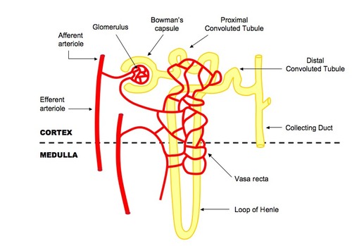
The nephron is the yellow tubule in the diagram above. It starts in the cortex with a cup-shaped structure called the Bowman’s capsule. This cup contains a tiny knot of capillaries called the glomerulus. The Bowman’s capsule empties into the second region of the nephron which is called the proximal convoluted tubule. The tubule then descends into the medulla and out again in a region called the Loop of Henle. There is then a second convoluted region called the distal convoluted tubule before the nephron empties into a tube called a collecting duct. The collecting ducts carry urine down into the renal pelvis and into the ureter.
Stages in the Production of Urine
1) Ultrafiltration
Blood is filtered in the kidney under high pressure, a process called ultrafiltration. Filtration is a way of separating a mixture of chemicals based on the size of the particles and this is exactly what happens to the blood in the kidney. Red blood cells, white blood cells and platelets are all too large to cross the filtration barrier. Blood plasma (with the exception of large plasma proteins) is filtered from the blood forming a liquid called glomerular filtrate. The kidneys produce about 180 litres of glomerular filtrate per day.
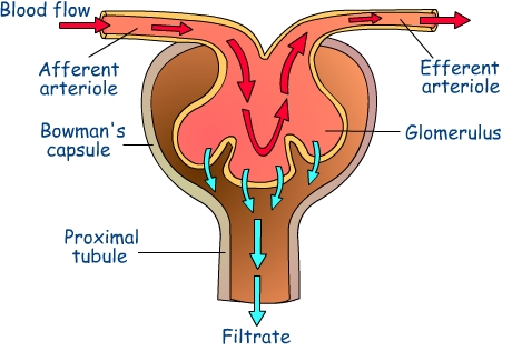
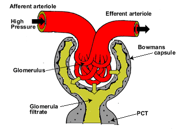
Ultrafiltration happens in the glomerulus and the glomerular filtrate (GF) passes into the Bowmans capsule. The high pressure is generated by the blood vessel that takes blood into the glomerulus (afferent arteriole) being much wider than the blood vessel that takes blood out of the glomerulus (efferent arteriole). The plasma of blood (minus the large plasma proteins) is squeezed out of the very leaky capillaries in the glomerulus and into the first part of the nephron.
What’s in Glomerular Filtrate?
- water
- glucose
- amino acids
- salts
- urea
As well as containing urea, water and salts, glomerular filtrate also contains many useful molecules for the body (glucose and amino acids for example) so these have to be collected back into the blood in the second stage…..
2) Selective Reabsorption
The useful substances in the glomerular filtrate are reabsorbed back into the blood. This can be by osmosis (for water) or by active transport (glucose and amino acids).
All of the glucose and all of the amino acids in the GF are reabsorbed in the proximal convoluted tubule by active transport. Remember active transport can pump substances against the concentration gradient using energy from respiration. Almost all the water in GF is reabsorbed by osmosis in the proximal tubule too.
So that leaves the question, what is the rest of the nephron doing…?
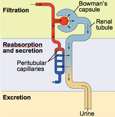

Well this is where it gets much more complicated…… Extra urea and salts can be secreted into the nephron at certain points along the tubule. The Loop of Henle allows the body to produce a urine that is much more concentrated than the blood plasma. And much of the distal tubules and collecting ducts are used for the second function of the kidney: homeostasis.
But you will have to wait until my next post to find out how the kidney fulfils this crucial second function… Please add comments or questions to this post – I really value your feedback… Tell me what is unclear and do ask questions….
Homeostasis: Grade 9 Understanding for IGCSE Biology 2.81, 2.82
Homeostasis is one of the more difficult topics for students to understand in the iGCSE specification. I have posted already about the skin and its role in thermoregulation so I suggest you read that post again to get the details….
https://pmgbiology.wordpress.com/2014/05/29/skin-a-understanding-for-igcse-biology/
In this post, I am going to try to explain the concept of homeostasis in much more general terms, then in later posts, look at the two examples mentioned in the syllabus. Here goes….
Homeostasis
Homeostasis is one of the life characteristics shared by all organisms. Living things all inhabit a world in which the external environment changes from hour to hour, from day to day, from month to month. Even organisms living in the most stable aquatic environments may be subject to changing oxygen concentrations, changing water pH, changing light intensities and so on. This changing external environment poses a challenge for life since how can life processes operate at optimal levels in all these differing conditions. Life has solved this by allowing organisms to keep their internal environments much more constant than the ever-fluctuating external environment.
A definition to learn:
“Homeostasis is the set of processes occurring in an organism to maintain a constant internal environment”
Examples of Homeostasis in Humans
A whole variety of factors are maintained at constant values in the body by homeostasis. For example (there are many more….):
- Blood pH
- Blood temperature
- Blood dilution
- Blood oxygen concentration
- Blood carbon dioxide concentration
- Blood glucose concentration
- Blood pressure
This introduces the first area of common confusion in students’ exam answers. For some reason many students think that homeostasis is a word for the maintenance of body temperature in humans. I hope you can see it is a much more general term than that.
But…. the systems that maintain a constant body temperature in endothermic animals are one example of homeostasis. In fact this example (thermoregulation) is one of the two from the list above that you need to understand for your exam. The other one you might be asked about is osmoregulation (the maintenance of a constant dilution of the blood).
All homeostatic control systems have some common features. The variable that is going to be regulated needs to be measured somewhere in the body. A change in this variable is called a stimulus and is measured by a cell called a receptor. The measured value needs to be compared with a “set value” and this is done by an integrating centre that then controls an effector. The effector is an organ that can bring about a response. But what kind of response do you want in the process of negative feedback?

A common process involved in homeostasis is negative feedback. This is quite tricky to define but in fact it is a really simple idea……. If you want things to stay the same, any change must be corrected. That’s negative feedback in a nutshell.
For example a school might want students walking round the campus at a sensible speed: not to fast to knock people over, not to slow or people are late for lessons… Imagine a particular group of children who start to run around the place, causing mayhem and injuries to fellow students. Well this will first be detected by the system. There may be a particular teacher who comes out and sees the students running, the school nurse might report an increase in cuts and bruises. However it happens, a change in the system (a stimulus) is detected. There will be an integrating centre in this control system too, probably in the form of a stern deputy head. She will compare the measured speed to her own “set value” of how fast students should move. And she will initiate a response: probably a loud telling off to the entire school in assembly, lots of dire warnings about future conduct and an after school detention for all the rule breakers. The net response of this will be that students will start moving slower around the school…. Eventually of course people will start moving too slowly and will be late for lessons. How do you think the system will react to this new stimulus? This process where the response tends to reduce the stimulus is called negative feedback.
To give you a biological example in thermoregulation, look at the diagram below:

If the body temperature goes up, the system responds by lowering the body temperature.
If the body temperature drops, the system responds by raising body temperature.
Both examples of negative feedback!

The end result of negative feedback is that the value will fluctuate around the set value (see this graph also showing the effect of negative feedback in thermoregulation)
Any questions, problems, queries: please comment using the box below the post or tweet me @Paul_Gillam and I will do my best to help…….

Nerve Cells and Synapses: Grade 9 Understanding for IGCSE Biology 2.88 2.89
There is very little in the iGCSE specification about nerve cells and synapses. This is a shame since neuroscience is going to be one of the massive growth areas in Biology in the 21st century. There is a syllabus point about reflex acs and I draw your attention to this blog post about that: https://pmgbiology.wordpress.com/2014/04/22/a-simple-reflex-arc/
But in this new post I am going to give you a tiny bit more detail about the types of nerve cells (neurones) that you might encounter, together with an explanation about the most important component of the nervous systems: the chemical synapse.
Neurones are the cells in the nervous system that are adapted to send nerve impulses. You won’t fully understand what the nerve impulse is until year 13 but it is correct so that it is a temporary electrical event that can be transmitted over large distances within a cell with no loss of signal strength. The upshot of this is that neurones can be very long indeed…..
There are three basic types of neurone that are grouped according to their function:
Motor neurones (efferent neurones) take nerve impulses from the CNS to skeletal muscle causing it to contract
Sensory neurones (afferent neurones) take nerve impulses from sensory receptors into the CNS
Relay (or sometimes Inter) neurones are found within the CNS and basically link sensory to motor neurones.
These three types of neurone also have different structures although many features are shared….

This is a diagram of a generalised motor neurone: I know it is a motor neurone since the cell body is at one end of the cell. The cell body contains the nucleus, most of the cytoplasm and many organelles. Structures that carry a nerve impulse towards the cell body are called dendrites (if there are lots of them) and a dendron if there is only one. The axon is the long thin projection of the cell that takes the nerve impulse away from the cell body. The axon will finish with a collection of nerve endings or synapses.

Neurones can only send nerve impulses in one direction. In the diagram above these two cells can only send impulses from left to right as shown. This is due to the nature of the junction between the cells, the synapse (see later on….)

The diagram above shows a sensory neurone. You can tell this because it has receptors at one end collecting sensory information to take to the CNS. The position of the cell body is also different in sensory neurones: in all sensory neurones the cell body is off at right angles to the axon/dendron.
You can see from the diagrams that motor and sensory neurones tend to be surrounded by a myelin sheath. Myelin is a type of lipid that acts as an insulator, speeding up the nerve impulse from around 0.5m/s in unmyelinated neurones to about 100 m/s in the fastest myelinated ones. The myelin sheath is made from a whole load of cells (glial cells) but there are gaps between glial cells called nodes of Ranvier. These will become important in Y12/13 when you study how the impulse manages to travel so fast in a myelinated neurone.
Relay neurones, also known as interneurones, have a much simpler structure. They are only found in the CNS, almost always unmyelinated and have their cell body in the centre of the cell.

The diagram above shows the three types of neurone and indeed how they are linked up in a simple reflex arc. The artist hasn’t really shown the interneurone structure very well, but it was the best I could find just now…..
Nerve cells are linked together (and indeed linked to muscle cells) by structures known as synapses. There are a lot of synapses in your nervous system. The human brain contains around 100 billion neurones and each neurone is linked by synapses to around 1000 other cells: a grand total of 100 trillion synapses. 100 000 000 000 000 is a big number.


The big idea with synapses is that the two neurones do not actually touch. There is a tiny gap called the synaptic cleft between the cells. The nerve impulse does not cross this tiny gap as an electrical event but instead there are chemicals called neurotransmitters that diffuse across the synaptic cleft.
The nerve impulse arrives at the axon terminal of the presynaptic neurone. Inside this swelling are thousands of tiny membrane packets called vesicles, each one packed with a million or so molecules of neurotransmitter. When the impulse arrives at the terminal, a few hundred of these vesicles are stimulated to move towards and then fuse with the cell membrane, releasing the neurotransmitter into the synaptic cleft. The neurotransmitter will diffuse rapidly across the gap and when it reaches the post-synaptic membrane, it binds to specific receptor molecules embedded in the post-synaptic membrane. The binding of the neurotransmitter to the receptor often causes a new nerve impulse to form in the post-synaptic cell.
These chemical synapses are really beautiful things. They ensure the nerve impulse can only cross the synapse in one direction (can you see why?) and also they are infinitely flexible. They can be strengthened and weakened, their effects can be added together and when this is all put together, complex behaviour can emerge. I am going to exhibit some complex behaviour now by choosing to take my dogs for a walk… And it all happened due to synapses in my brain!
Adrenaline: Grade 9 Understanding for IGCSE Biology 2.94
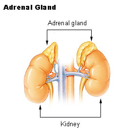
Adrenaline is a hormone produced in the adrenal glands which are found on top of the kidneys in the abdomen.
A hormone is “a chemical released by a specialised gland called an endocrine gland into the bloodstream. The hormone travels around the body in the blood plasma and then causes an effect elsewhere in the body by binding to receptors found on certain target cells”.
You should know some other examples of hormones – testosterone, oestrogen, progesterone, ADH – to name a few. Please learn this definition too: it would be wonderful if you got a 3 mark question asking you to define a hormone….

There are many cells in the body that contain receptors for adrenaline. This allows the hormone to exert an effect on a wide variety of tissues. For example there are adrenaline receptors in the pacemaker of the heart and adrenaline will cause the heart to beat faster (more beats per minute) and also with more force.
When is adrenaline released by the adrenal glands into the blood?
Adrenaline is secreted into the blood in times of danger or stress. It prepares the body to either run away from the danger or indeed to battle against it. For this reason, adrenaline is often described as a “fight or flight” hormone.
What are some of the effects of adrenaline?
Target Tissue Effect
Heart Increase in heart rate, increase in cardiac output
Lungs Bronchioles dilate (widen)
Muscles Arteries in muscle dilate to allow more blood to flow to muscles
Skin/Digestive system Arteries in skin/digestive system constrict so less blood flows
Liver Liver breaks down glycogen into glucose to raise blood glucose conc.
Iris Radial muscles in iris contract causing pupil dilation
The overall effect is that the skeletal muscles are supplied with more oxygen and more glucose so they can respire aerobically. This allows the muscle to contract more efficiently.

Cardiac cycle and the Human Heart: Grade 9 Understanding for IGCSE Biology 2.65 2.66
The human heart is an organ found in the middle of the thorax. It is made from a specialised type of muscle called cardiac muscle and acts as a pump to push blood around the two circulatory systems. In fact it is better to think of the heart as two separate pumps, one for the pulmonary circulatory system to the lungs and one for the systemic system that supplies blood to all the other body organs.
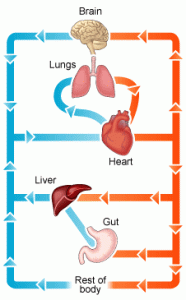
The diagram above shows in a simplified way this double circulatory system. The lungs are supplied with deoxygenated blood direct from the heart in the pulmonary artery but when the blood has passed through the capillaries in the lungs, it travels back to the heart in the pulmonary vein before being pumped in the systemic system around the body. Although cardiac muscle looks similar to the muscle that attaches to bones and moves the skeleton, it differs functionally in one important way. Cardiac muscle is myogenic: this means that the muscle fibres will contract without the need for a nerve impulse from the brain to initiate the contraction. Incidentally this is why a heart transplant is a possible surgical procedure. A transplanted heart will beat happily in the new body even though all the nerves going to the heart will have been cut in the surgery. You cannot have a biceps transplant at the moment because the transplanted biceps muscle would not do anything in the new patient. For the transplanted biceps to contract, the millions of individual neurones going to it would need linking up individually and this is not possible.

This is a simplified diagram showing the structure of the heart. You can see that the left and right sides of the heart are completely separate from each other. This is essential because the right side of the heart contains deoxygenated blood and the left side oxygenated blood. There are four chambers in the heart: two small atria at the top, and two larger ventricles at the bottom. The atria collect blood from the veins, the ventricles pump blood out of the heart into arteries.
There are four sets of valves in the heart: the easiest way to remember where they are is to think that blood has to pass through a set of valves as it leaves each chamber. Valves allow blood to flow through them in one direction only. The AV valves stop blood going back from the ventricle into the atrium when the ventricle contracts, the aortic and pulmonary valves (not labelled on the diagram above for some reason….) prevent blood falling back into the ventricles in between heart beats.
You also need to know the four main blood vessels that are attached to the heart. The vena cava is the largest vein in the body and carries deoxygenated blood from the organs of the body into the right atrium. The pulmonary artery comes out of the right ventricle and pumps this deoxygenated blood to the lungs. Oxygenated blood returns from the lungs to the left atrium in the pulmonary veins, passes into the left ventricle and is then pumped out of the heart in the aorta, the biggest artery in the body.

NB I strongly suggest you do not use the terms bicuspid, mitral or tricuspid valve to label the valves between the atria and ventricles… In an exam it is easy to get these similar words muddled, so I would always call the valve between the left atrium and left ventricle the left atrio-ventricular valve (never bicuspid or mitral valve) and I would always call the valve between the right atrium and right ventricle the right atrio-ventricular valve (never tricuspid). I know many of you will ignore this advice but it’s good to get it off my chest……
I’ve just spent 45 minutes trying to find a good video on heart structure to put into the post but without success…. If anyone knows a really good YouTube clip (must be under 5 minutes) please add a link as a comment at the foot of this post.
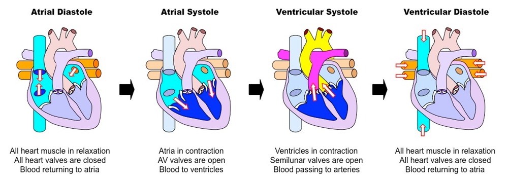
Events of the Cardiac Cycle
This is a topic that requires some simplification as it is easy to get confused. The Cardiac Cycle is simply a word for the sequence of events that happen in a heart beat. At the simplest level, the cardiac cycle consists of three phases:
1. Diastole. During this stage the cardiac muscle is relaxed (the heart is between beats) and blood can enter the atria and then fall into the ventricles through the open AV valves.
2. Atrial Systole. This stage in when the cardiac muscle in the atria contract, increasing the atrial pressure and pushing blood down into the ventricles. There is a small region in the wall of the right atrium called the sino-atrial node (or pacemaker) which initiates each heart beat.
3. Ventricular Systole. After a short delay, the cardiac muscle in the ventricles contracts. This increases the blood pressure in the ventricle which in turn causes the AV valve to close, and the aortic or pulmonary semilunar valves to open. As these valves at the exit of the ventricle open, blood gets pushed into the arteries and out of the heart.
Look at the diagram above and make sure you understand the meaning of these three terms.
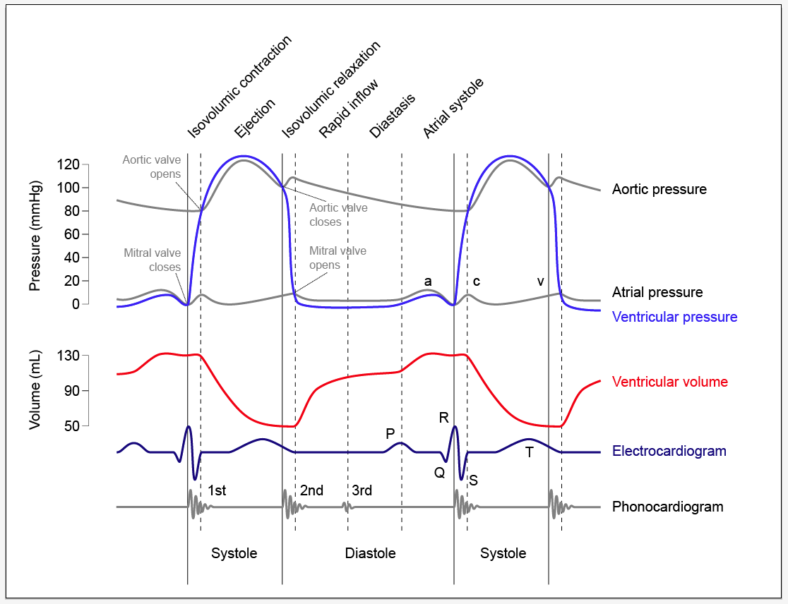
The diagram below is much more complex and perhaps you should not worry too much about it….. The important bit is to understand when the valves in the heart open and when they close. It is quite simple really although you would be amazed how confused people can get….
The opening and closing of a valve is not “controlled” in any meaningful way in the heart. The valve has a structure that will only allow it to open in one direction. Let’s consider the AV valves. These can open to allow blood to pass from the atria into the ventricles during atrial systole but will close during ventricular systole to stop the blood flowing back where it came from.
The AV valve will be open whenever the blood pressure in the atria is greater than in the ventricle.
The AV valve will be closed whenever the blood pressure in the ventricle is greater than in the atrium.
Look at the graph of pressure changes below. You can see the mitral valve (I hate that name) is closed as soon as the ventricular pressure exceeds the atrial pressure and it opens again during ventricular diastole as the pressure in the ventricle drops.

Blood part 2 White Blood Cells – Grade 9 Understanding for GCSE Biology 2.59 2.62 2.63B
The previous post looked at the structure and function of red blood cells and plasma. Now it is time to turn our attention to the rather more complex topic of white blood cells….. This is a topic in which the complexity can put people off but I am deliberately going to keep things simple (I hope!). If you are thinking about revision for GCSE, don’t worry about anything more complicated than in this post.

There are many types of white blood cell found in blood. But let’s keep things simple…. You need to understand the role of lymphocytes and phagocytes in defending the body against pathogens.
A pathogen is defined as “a microorganism that can cause a disease” and pathogens may be bacteria, viruses, protistans or fungi. Can you give me an example of an infectious disease caused by each class of pathogen?
The structure of these two classes of white blood cell is important. The commonest phagocytes in blood are called neutrophils and they are easily recognised by their irregular shaped nucleus and cytoplasm packed full of granules. Lymphocytes are much smaller white cells and are identifiable by their clear cytoplasm and large spherical nucleus that takes up 90% of the volume of the cell.


So now we should look at how these two types of white blood cells defend the body against pathogens. Remember that the account on this post is an over-simplification of what is in reality an extremely complex process.
Let’s start with a phagocyte. These large cells are able to engulf invading pathogens in the blood and tissue fluid by a process called phagocytosis.

The phagocyte pushes out projections of its cytoplasm around the clump of bacteria. These projections are called pseudopodia and when they meet, the cell membrane of the phagocyte fuses together leaving the bacteria enclosed in a tiny membrane packet called a vesicle inside the cytoplasm. The phagocyte then fuses other vesicles that contain powerful digestive enzymes with the vesicle with the bacteria in, leading to the death and destruction of the bacteria. Simple.
The problem for phagocytes is this: how do they know what to engulf and destroy? This is where lymphocytes come in. One class of lymphocyte is able to secrete small soluble proteins called antibodies into the blood. Antibodies are specific to a particular surface marker on the invading pathogen and bind to it because the shape of the antibody and the shape of the surface marker are complimentary.

Now people always get confused between antibodies (the small soluble Y-shaped proteins secreted by lymphocytes) and antigens (the surface markers on the invading pathogen). Make sure you are completely clear on the difference in meaning of these two words….
 This diagram shows antibodies (green) binding to surface markers (antigens) on a bacterial cell.
Antibodies produced by lymphocytes will coat the invading pathogen by binding to antigens on its surface. One effect of this is that phagocytes are stimulated to engulf the antibody-coated organism.
There are many different types of lymphocyte and not all can produce antibodies. Another important function of lymphocytes is to kill your own body cells when they are corrupted, either by the presence of a virus or by becoming cancerous.
Finally, can I draw your attention to two previous posts linked to this one. The first is on the role of platelets in blood clotting, the second on the difficult topic of immunity and how lymphocytes are responsible for giving you lifelong protection against certain infectious diseases.
https://pmgbiology.wordpress.com/2014/04/07/immunity-a-understanding-for-biology-igcse/
As always, please ask me questions either via the comment section below the post or with a tweet…. I will do my best to respond to any questions from anyone who is bothered to read my posts!
Blood part 1 Plasma and RBCs: Grade 9 Understanding for IGCSE Biology 2.59, 2.60, 2.61
Blood is a tissue in the body that plays a variety of roles in transport and in defending the body against disease. It is an unusual tissue since it is a liquid, with many different kinds of cells suspended in a watery solution called plasma. 
Plasma makes up 55% of the volume of blood and is a solution of many different chemicals in water. For example, the plasma contains dissolved glucose, amino acids and other products of digestion from the intestines. It also transports the waste molecule urea from the liver where it is made to the kidney where it is excreted. Blood plasma contains dissolved carbon dioxide, mostly in the form of hydrogencarbonate ions. Many hormones (for example testosterone, ADH, adrenalin) are transported in the blood plasma and because the plasma is mostly water, it provides a good way of moving heat around the body from respiring muscles to the skin where it can be lost.
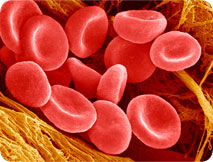
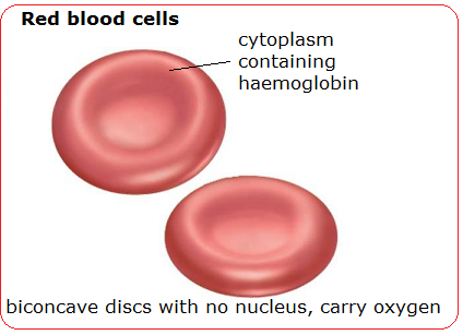
The most common cell in blood are the red blood cells (or erythrocytes). These tiny cells are adapted for the transport of oxygen. Each red blood cell contains around 270 million molecules of a transport protein, haemoglobin. Each molecule of haemoglobin can bind up to four molecules of oxygen in the lungs and then unload the oxygen when the red blood cell passes through a capillary in an actively respiring tissue.

(Don’t worry too much about the structure of the protein – this is A level stuff really…. Just remember haemoglobin is a transport protein for oxygen found in red blood cells)
As well as being packed full of haemoglobin molecules, red blood cells have other adaptations for transporting oxygen. Red blood cells lose their nucleus during their development as this allows more haemoglobin to be packed into each cell. Having no nucleus means the red blood cell cannot divide nor repair damage to its structure. This is why each red blood cell only lives for 100-120 days in the body.
Red blood cells have a characteristic shape. It is called a biconcave disc and they have an especially flexible shape. Remember that a capillary is actually smaller in diameter than a red blood cell, so the cells have to squeeze through capillaries in single file…..

Blood vessels – Grade 9 Understanding for IGCSE Biology 2.68
In this post, I will look at the structure and function of the three main types of blood vessel in the human circulatory system. Although this is not the most difficult topic, there are a few things that can catch out even A* GCSE students in the heat of an exam.
Arteries are the blood vessels that take blood away from the heart. Because the blood is coming straight from the ventricles of the heart, it will be at a high blood pressure and will flow in pulses. This means that arteries need a thick wall to withstand this high blood pressure. All arteries apart from one carry oxygenated blood. Can you remember which artery is the exception to this rule?

The artery wall has a narrow lumen (the space where the blood flows) as this helps to maintain the high blood pressure within. There are also many elastic fibres in the middle tissue (tunica media) of the artery wall. This elastic tissue is important because the blood flows in pulses. The artery wall needs to stretch as the pulse of blood passes and the elastic recoil of the wall helps to push the blood along in between heart beats.
The tunica media also contains a lot of smooth muscle. Why do arteries need muscle in their walls? When this muscle contracts it narrows the lumen of the vessel. This will increase the blood pressure and so one reason for muscle in arteries is to regulate the blood pressure. But there is something more… Arteries carry blood into the organs of the body and the pattern of blood flow to different organs can vary depending on the conditions. For example, when you are running, you need more blood to go to your skeletal muscles (to carry oxygen for respiration and to remove heat and carbon dioxide) and less to go to the digestive system. This is brought about by the smooth muscle in the artery taking blood to the intestines and stomach contracting so that less blood can flow through the vessel. The smooth muscle in the arteries in the exercising muscles will relax so that more blood can pass. This shift in the pattern of blood flow is the second key significance of arteries having lots of muscle in the walls.

Veins have the same tissues in their walls as arteries but they are much thinner. The blood is flowing at a much lower pressure in veins as all the pressure from the heart has been lost in the extensive capillary beds in the tissues. Veins return blood to the heart and all bar one (the pulmonary vein) contain deoxygenated blood. As there is low blood pressure in veins, this can cause problems moving blood back to the heart especially when against gravity. Veins contain valves which only allow blood through in one direction thus preventing the blood falling back. The thin walls of veins also mean that they can be compressed by the action of skeletal muscles. When the muscles that move the skeleton contract, they can squeeze on veins and help to return blood to the heart.

Capillaries are the smallest of the three types of blood vessel. They are found in the tissues throughout the body and are beautifully adapted to ensure the exchange of materials between the cells of the body and the blood. The lumen of a capillary is less than the width of a red blood cell and so red blood cells pass through capillaries in single file and only be squeezing along. This ensures the speed of blood flow in capillaries is very very slow.
The lining of a capillary is made up of a single layer of cells called the endothelium. Arteries and Veins have an endothelium too but in the capillary the endothelial cells have gaps between them called pores. This allows the fluid component of blood and various white blood cells to leak out of capillaries to form tissue fluid. This leaky nature of capillaries is very important as it provides the fluid that bathes the tissues of the body.
