
Tagged: chromosome
Chromosomes and Sex: Grade 9 Understanding for IGCSE Biology3.26 3.27
Having spent the last day or two writing material about one of the hardest topics in the IGCSE Biology specification (DNA and Protein Synthesis), I am going to write today about something much simpler. You need to understand how the sex of a human is determined at the moment of fertilisation. But this is a topic which can confuse students so I am going to try to explain it for you as best I can.
The sex of a human (whether male or female) is determined by the 23rd pair of chromosomes. Please remember that just because humans determine their sex this way, this doesn’t mean that other species have to be the same. In fact other species use a variety of ways to ensure the correct proportion of male and offspring are born.

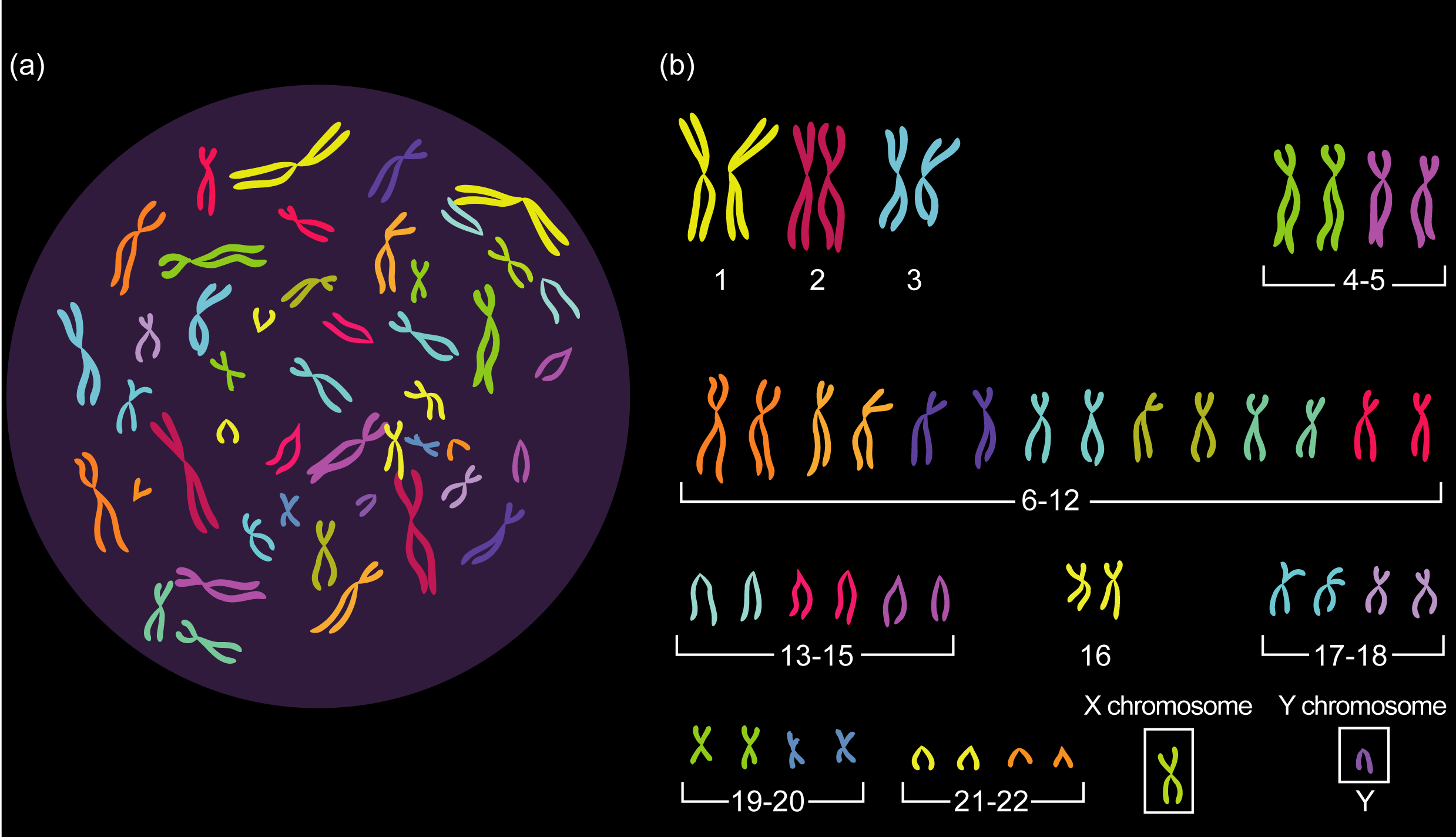
As you can see from the picture above, the 23rd pair of chromosomes in humans are called the sex chromosomes. The person whose chromosomes are shown above is male because he has one X and one Y chromosome in his 23rd pair. If we looked at a picture of a human female set of chromosomes, pairs 1 to 22 would be exactly as above, but the 23rd pair would be different. There would be two large X chromosomes rather than one large X and one tiny Y chromosome as shown above.
So a human female has XX as her 23rd pair of chromosomes, a human male has XY as his 23rd pair.
Gametes (Sperm and Egg cells) are made in a process called Meiosis. Remember that meiosis produces daughter cells that are haploid (this means they only have one member of each pair of chromosomes and so half the genetic material)
When a female cell undergoes meiosis in her ovary, the daughter cells produced (egg cells) will contain one of each of the 23 pairs of chromosomes. For the 23rd pair this will always be an X chromosome since both chromosomes in the 23rd pair are X chromosomes.
When a male cell undergoes meiosis in the testis, the daughter cells produced (sperm cells) will contain one of each of the 22 pairs of chromosomes exactly as above. But the 23 pair are different to each other and so half the sperm cells will contain an X chromosome as the 23rd chromosome and half the sperm cells will contain a Y chromosome as the 23rd chromosome.

If you understand the picture above, you understand sex determination in humans. You also need to be able to draw a genetic diagram to show this.
Phenotype: Mum Dad
23rd pair: XX XY
Gametes: X ½X ½Y
Fertilisation:

Offspring 23rd pair of chromosomes: ½ XX and ½ XY
Offspring phenotypes: ½ female and ½ male

Cell Division part 3: Grade 9 Understanding of Meiosis for IGCSE 3.30
In sexually reproducing organisms two types of cell division are needed. One is for the processes of growth, repair and asexual reproduction and it is called mitosis. Mitosis produces daughter cells that are diploid and genetically identical to the parent cell.
But when the organism wants to make gametes a different mechanism is needed. Gametes are not diploid like all the other body cells, but instead they only have one member of each homologous pair of chromosomes. In order to make a haploid daughter cell, a second type of cell division, meiosis, is needed.

You can see in the diagram above some of the key differences between mitosis and meiosis. Both start with diploid cells (2n) but whereas mitosis involves one round of division and produces two identical diploid daughter cells, meiosis is different. Meiosis has two rounds of division, called Meiosis I and Meiosis II. This results in four daughter cells and you can see that they are all haploid (n) cells. These cells develop into gametes (sperm and egg cells in humans) and so when they fuse together in fertilisation, the diploid number is restored.

Gametes are all genetically different to each other
Meiosis does not just produce haploid daughter cells. It also introduces genetic variation into the daughter cells so each is genetically unique. This means that random fertilisation will produce offspring that are all genetically different to either parent. How does this genetic variation in gametes come about?
Well to answer that, you need to understand a little bit more about how the chromosomes behave during meiosis. I am not going to talk through all the various stages of meiosis (life is too short and you can read the diagram below) but there is a key event that happens in meiosis that never happens in mitosis…..
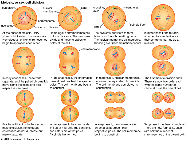
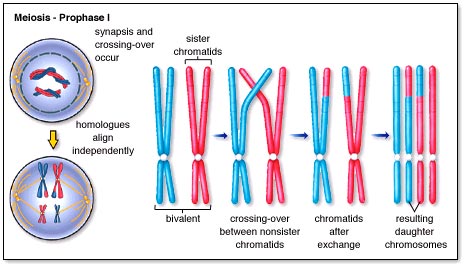
It happens in prophase of the first meiotic division and is called synapsis. As the nuclear membrane is degenerating, the two members of a homologous pair of chromosomes line up alongside each other to form a structure called a bivalent (or tetrad)
In the first meiotic division, the two members of the homologous pair are pulled apart and separated. Because these bivalents attach and assort independently of each other, this means that this random assortment can produce many different gametes. A human cell with 23 pairs of chromosomes can produce 2^23 possible gametes just by random assortment.
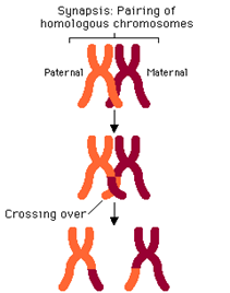
But there is a second process called crossing over that happens during prophase 1 when the bivalents are formed. As you can see in the diagrams, small sections of chromatid can be swapped between the chromatids of one chromosome and with its homologous partner. This ensures that when the individual chromatids are separated in meiosis 2, each is different to each other. This multiplies up the genetic variation by several orders of magnitude. (see diagram below)
Now that is more detailed than you will need in an iGCSE exam, but it is good to understand where the genetic variation in gametes comes from. Let’s finish with something more simple – the differences between mitosis and meiosis.
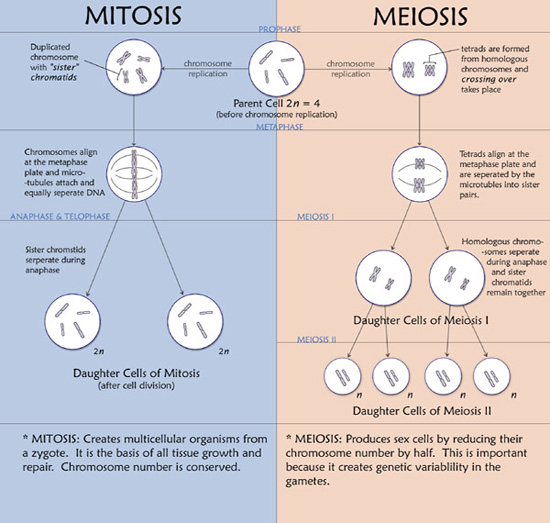
One final point: please learn the spellings of these two types of cell division. Spelling is only penalised in exams when the meaning is lost and any intermediate spelling (e.g. meitosis or miosis) has no meaning! So if you are one of those people who finds spelling difficult, find a way of learning mitosis (produces identical diploid daughter cells and is used in growth) compared to meiosis (produces genetically different haploid cells and is used to make gametes)
Cell Division part 2: Grade 9 Understanding of Mitosis for IGCSE 3.28 3.29
I thought I would make a video to explain the details of mitosis rather than typing up a blog post.
I would welcome any feedback on either of these videos. Do you find them useful? Do you prefer written blog posts? Leave a comment below if you want to let me know…..
Cell division video: a revision video for DNA structure and chromosomes
This is a summary video that might help those of you still struggling to get to grips with chromosomes and genes. I apologise for the terribly amateur production values on the video but hope the biological content at least might be useful….
Cell Division part I: Grade 9 Understanding for IGCSE 3.15, 3.28, 3.29
At the beginning of March each year, I get my Y11 classes to draw up a list of topics they want to go through again in revision. Cell Division is always there and it is not difficult to see why. Mitosis doesn’t make any sense unless you understand the concept of homologous pairs of chromosomes and I think you already know that very few iGCSE students do…… (Please see the various posts and videos on the blog on this topic before attempting to understand mitosis)
But there is actually very little to fear in the topic of cell division. If your teacher has told you about the various stages of mitosis that’s fine but you will not be asked to recall them in the exam, at least not if you are studying EdExcel iGCSE. So in this post I am going to try to focus on the key bits of understanding you need rather than bombarding you with unnecessary details.
1 Chromosomes come in pairs
This is the main idea you need before you start. In almost all sexually-reproducing organisms the cells are DIPLOID. This means that however many different sized chromosomes they have, in each cell there will be pairs of chromosomes (called homologous pairs)
So human cells contain 23 pairs of chromosomes (46 in total)
Remember that the number 46 only applies to humans. Other species have very different numbers of chromosomes in each cell (see table below)
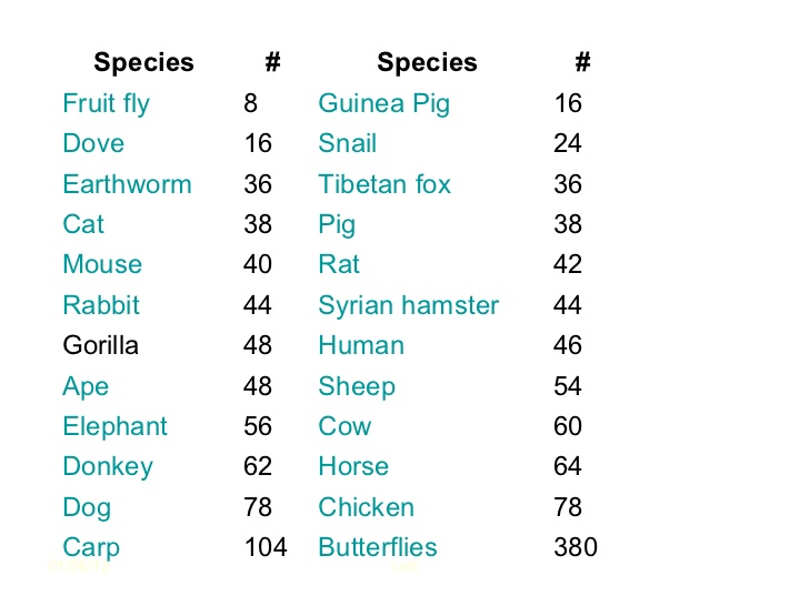
So doves have 8 pairs of chromosomes, dogs have 39 pairs of chromosomes, rats 21 pairs of chromosomes. The important point is not how many pairs each organism has but that they all have chromosomes that come in pairs!
The chromosomes any individual possesses is determined at the moment of fertilisation. Sperm and Egg cells (gametes) do not have pairs of chromosomes. They are the only cells in the body that are not diploid. Gametes only have one member of each pair of chromosomes. Cells which only have one member of each pair of chromosomes are called HAPLOID cells.
So every cell in the body is diploid and genetically identical apart from the gametes which are haploid.
2 Organisms that reproduce sexually need two different types of cell division
The fertilised egg (zygote) is a diploid cell. It has pairs of chromosomes that originate one from each parent via the gametes. Every cell division in growth and development of the embryo and foetus until birth, every cell division in growth and repair after birth always produces two genetically identical and diploid cells from the one original cell. This cell division that produces genetically identical diploid cells is called Mitosis.
Gametes (sperm and egg cells) need to be made by a different process. If gametes were diploid then there would be a doubling of the chromosome number every generation and that clearly wouldn’t do. So a different way of dividing the nucleus has evolved. It doesn’t produce genetically identical diploid cells but produces gametes that are haploid and genetically unique. This process is called Meiosis and is only used in the production of gametes.
3 Mitosis is involved in growth, repair, asexual reproduction and cloning
Any process in the body in which the outcome required is the production of genetically identical diploid cells will use mitosis. (It is not too complicated an idea to see that if you don’t need to make gametes and fuse them together in fertilisation, you can just copy cells by mitosis over and over again. All the daughter cells will be exact copies of each other and diploid.
Now I know this post is not going to satisfy everyone. I know some of you will want to read about the cell cycle, prophase, metaphase, centrioles, spindle fibres and the condensation of chromosomes, chromatids being pulled apart etc. etc.) And just for you, I will write a post later today on the details of Mitosis….. But please remember that if you are using the blog to revise for exams, none of this second post is necessary and none of it will be tested in the Edexcel iGCSE paper. If you are doing revision, focus on the key understanding ideas discussed above. And as always, please leave a reply below to ask questions, comment or leave feedback – all comments welcome!

Chromosomes video – a short PMG summary of some important ideas for GCSE
This is my first attempt at making a short summary video as a follow up to lessons. I apologise for the poor sound in places – I hope the content makes sense. Please tweet or leave a reply with any questions.
Chromosomes: Grade 9 Understanding for IGCSE Biology 3.15 3.32
I hope everyone reading this blog knows the definition of a gene. It is one of the few things in the iGCSE course that it is worth learning by heart.
“A gene is a sequence of a DNA molecule that codes for a single protein“.
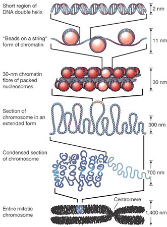
In human cells, every nucleus contains about 23,000 genes. Remember there is about 1.5m of DNA inside each nucleus. For most of the life-cycle of the cell, this DNA is in a tangled web called chromatin. Chromatin is DNA molecules loosely associated with some scaffolding proteins. The scaffolding proteins are shown in the second level down of this excellent diagram as “beads on a string”.
But this tangled web of DNA in chromatin poses a problem for the nucleus. For the cell to divide by mitosis, it is essential that the nucleus replicates into two identical nuclei, one for each new cell. The DNA molecules in the nucleus will make a copy of themselves by semi-conservative replication but how then can you ensure that each daughter nucleus gets exactly one copy of each DNA molecule if they are all tangled up….? This is where chromosomes come in!
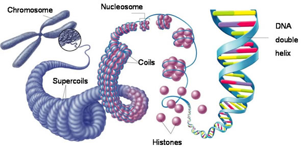
Each chromosome is a physical structure formed by supercoiling of the DNA round the scaffold proteins. The DNA coils, then folds back on itself, then coils again until each DNA molecule is so tightly coiled up that a visible chromosome appears in the nucleus. Chromosomes only become visible just before mitosis starts as for the rest of the time, the DNA is much more loosely coiled and so cannot be seen.
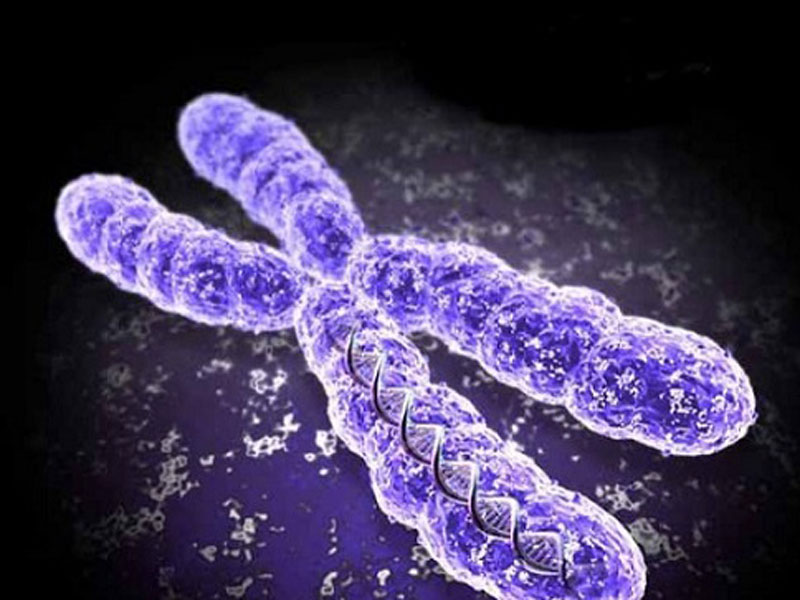
This also explains why each chromosome always looks X shaped. When chromosomes become visible the DNA has already replicated, so one chromosome is now made of two identical sister chromatids joined at a region called the centromere.

So the picture on the left shows a chromosome made as a single structure comprising one DNA molecule wrapped around the scaffold proteins. Then DNA replication occurs (in the S phase of the cell cycle) and now each chromosome is made of two identical chromatids joined at the centromere. Then the two chromatids are separated in mitosis and the chromosome returns to the structure it had at the start.
How many chromosomes are there in human cells?
The key idea here is that chromosomes are found in pairs in all body cells apart from gametes, These pairs of chromosomes (called homologous pairs) have exactly the same genes in the same locations on the chromosome. They are inherited one from each parent so one member of each pair will come from your father, one from your mother.
Different species have different numbers of pairs of chromosomes. For humans you should know that we have 23 pairs of chromosomes in the nucleus of every body cell (making a total of 46). Cells with chromosomes found in pairs are called diploid cells. Every cell in the body is diploid apart from the gametes. Gametes only have one member of each homologous pair and are called haploid cells.
Which of the following cells are diploid, which are haploid?
- Zygote
- Skin cell
- Sperm cell
- Liver cell
- Pollen grain
- Egg cell
If you are not sure, ask me by leaving a comment below….
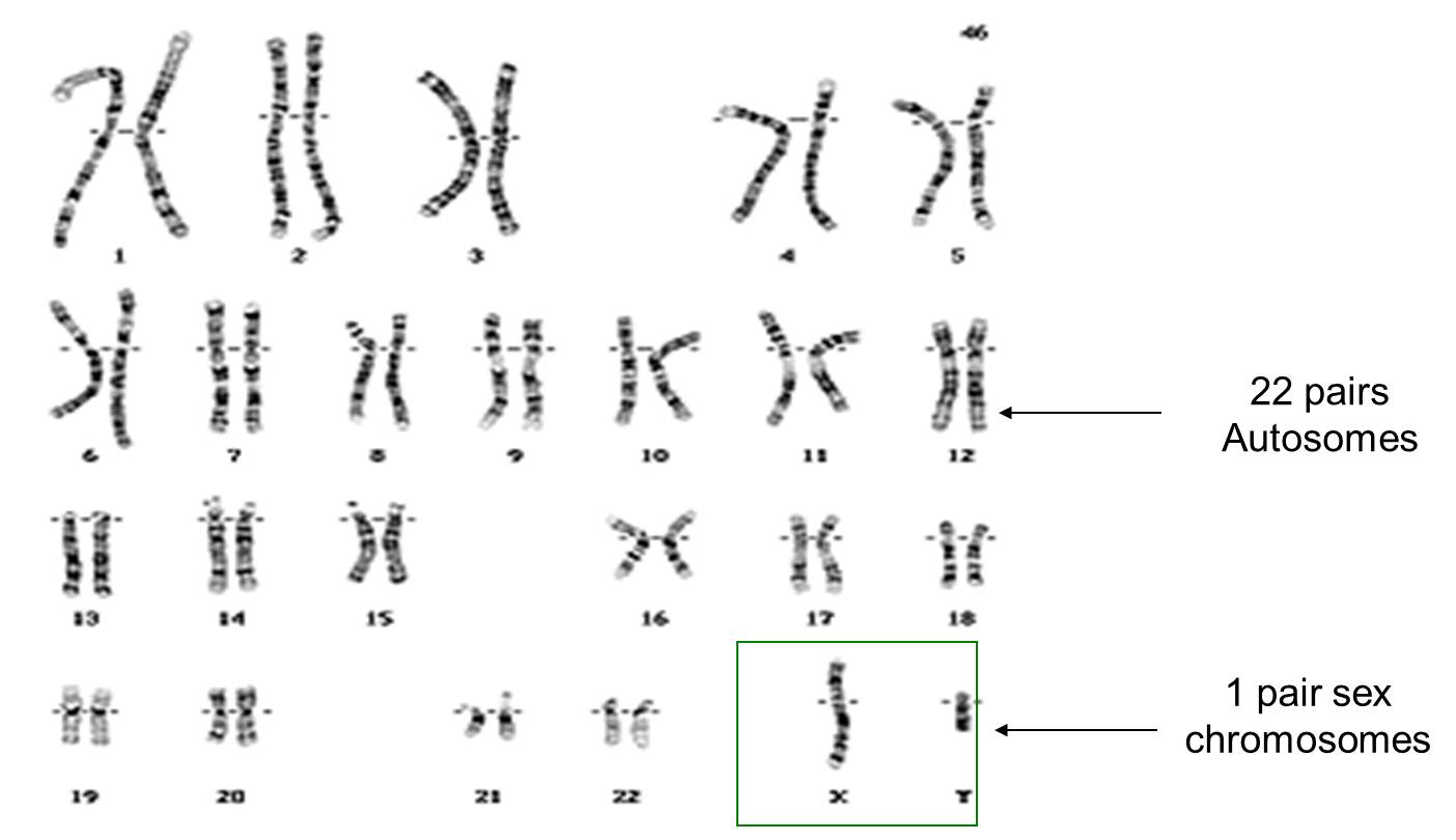
Finally for this post, chromosomes determine the sex of a human. You can see in the picture above that the 23 pairs of chromosomes can be divided into pairs 1 to 22 – these are called autosomes and play no role in determining your sex. But the 23rd pair of chromosomes are called the sex chromosomes. Males have one large X chromosome and one tiny Y chromosome as their 23rd pair whereas females have two large X chromosomes.
Gametes are haploid so only have one member of each pair. So when a man makes sperm cells (by meiosis) 50% of his sperm cells will contain his X chromosome, 50% his Y chromosome. A woman’s egg cell will always contain one X chromosome. (Why is this?) So I hope you can see that at the moment of fertilisation, the babies sex is determined depending on whether it is a Y-containing sperm cell that happens to fertilise the egg or an X-chromosome containing sperm… If the former, the baby is male, if the latter female.
I might explain this more fully in a post some other time….
Final thing for this post. If you have got to the end of this and understand everything in the text above, you are in a tiny minority of school students. Well done! This is a tricky topic and if you really understand chromosomes, you stand a chance of understanding cell division and genetics.
Mutation – Grade 9 Understanding for IGCSE Biology 3.34 3.37B
Mutations are changes in the DNA content of a cell. There are various ways the DNA of a cell could change and so mutations tend to be grouped into two main categories: chromosomal mutations and gene mutations.
Chromosomal mutation
This is a change in the number or length/arrangement of the chromosomes in the nucleus. For example, people with Down’s syndrome have an extra copy of chromosome 21 giving them three chromosome 21s as opposed to the normal two.
(How many chromosomes in total will a person with Down’s syndrome have in each cell?)
Chromosomal mutations are often found in tumour cells and so play a critical role in the development of various cancers.
Sometimes the number of chromosomes in a cell stays the same, but sections are deleted, duplicated or break off from one chromosome to attach elsewhere. If this happens, this too would be classed as a chromosomal mutation.


Gene mutation
Gene mutations happen to change the sequence of base pairs that make up a single gene. As you all know, the sequence of base pairs in a gene is a code that tells the cell the sequence of amino acids to be joined together to make a protein. A gene then is the sequence of DNA that codes for a single protein. If you alter the sequence of base pairs in the DNA by adding extra ones in, or deleting some or inverting them, this will alter the protein produced.
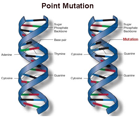
A point mutation is a change to just one base within the gene – it occurs at a single point on the DNA molecule.
Mutations can happen at any time and occur randomly whenever the DNA is replicated. But there are certain things that can increase the rate of mutation and so make harmful mutations more likely. A mutagen is an agent that increases the chance of a mutation occurring.
a) Radiation can act as a mutagen
Some parts of the electromagnetic spectrum can cause mutations when they hit DNA molecules or chromosomes. This is called ionising radiation and includes gamma rays, X rays and ultraviolet. You probably know that the dentist goes out of the room whenever they take an X ray to protect themselves from repeated exposure to X rays and you all certainly know of the link between UV exposure and incidence of skin cancer.
b) There are chemical mutagens as well
Some chemicals can make the rate of mutation increase. These are called chemical mutagens and a good example is the tar in tobacco smoke. Tar can cause cancers to form wherever the cigarette smoke comes into contact with cells and this is because tar is a mutagen. It makes mutations in the DNA much more likely and mutations are needed to turn a healthy cell into a cancer cell.
