
Category: Section 3: Reproduction and Inheritance
Hormones: Grade 9 Understanding for IGCSE Biology 2.94 2.95B
Hormones are defined as “chemicals produced in endocrine glands that are secreted into the bloodstream and cause an effect on target tissues elsewhere in the body”. They play a wide variety of roles in the healthy functioning and development of the body.

The iGCSE specification only really mentions a small number of hormones so these are the ones I will focus on in this post.
ADH (anti-diuretic hormone) (Separate Biologists only – not Combined Science)
ADH is secreted into the blood by an endocrine gland at the base of the brain called the Pituitary Gland. The stimulus for the release of ADH into the blood comes from the hypothalamus (a region of brain right next to the pituitary gland) when it detects that the blood plasma is becoming too concentrated. This might be caused by the body becoming dehydrated due to sweating. ADH travels round the body in the blood until it reaches its target tissue which are the cells that line the collecting ducts in the nephrons in the kidney. ADH increases the permeability of the connecting duct walls to water, thus meaning more water is reabsorbed by osmosis from the urine in the collecting duct and back into the blood. This results in a small volume of concentrated urine being produced.
Adrenaline
Adrenaline is secreted into the blood by the adrenal glands in situations of danger or stress.. The adrenals are found just above the two kidneys on the back of the body wall. Adrenaline secretion is controlled by nerve cells that come from the central nervous system. Adrenaline is often described as the “fight or flight” hormone as its effects are to prepare the body to defend itself or run away from danger. There are receptors for adrenaline in many target tissues in the body but some of the most significant effects of adrenaline are:
- affects the pacemaker cells in the heart causing an increase in heart rate
- shifts the pattern of blood flow into muscles, skin and away from the intestines and other internal organs
- decreases peristalsis in the gut
- causes pupils to dilate in the eye
- increases breathing rate in the lungs
- promotes the passing of urine from the bladder
Insulin
Insulin is a hormone made in the islets of Langerhans in the pancreas. It plays a vital role in the homeostatic control of the blood sugar concentration. The pancreas will secrete insulin into the blood when the blood glucose concentration gets too high. There are many cells in the body with insulin receptors but the main target tissue for insulin is the liver.
Insulin causes the liver (and muscle) cells to take glucose out of the blood and convert it into the storage polysaccharide glycogen. This results in a lowering of the blood glucose concentration: a good example of the importance of the principle of negative feedback in homeostasis
Testosterone
Testosterone is a steroid hormone made by cells in the testes of males. It is the main hormone of puberty in males resulting in the growth of the reproductive organs at puberty as well as the secondary sexual characteristics (pitch of voice lowering, muscle growth stimulated, body hair grows etc.)
Oestrogen
Oestrogen is a steroid hormone made by the cells in the ovary that surround the developing egg cell in the first half of the menstrual cycle. In puberty it causes the development of the female secondary sexual characteristics (breast growth, change in body shape, pubic hair etc.) but in the menstrual cycle, oestrogen has a variety of important effects. It stimulates the rebuilding of the uterine endometrium (or lining) to prepare the uterus for the implantation of an embryo. Oestrogen also affects the pituitary gland and can cause the spike in LH concentrations that trigger ovulation on day 14 of the cycle.
Progesterone
Progesterone is also made in the ovary but at a different time in the menstrual cycle. It is secreted by cells in the corpus luteum, a structure found from day 14 onwards after the egg has been released in ovulation. Progesterone has two main target tissues: it maintains the thickened lining of the endometrium in the uterus ready for implantation. Progesterone also causes the pituitary gland to stop secreting the hormones FSH and LH so a new cycle is never started. It is for this reason that progesterone can be used in women as a contraceptive pill.
FSH (Follicle-Stimulating Hormone (Separate Biologists only – not Combined Science)
FSH is a hormone released by the pituitary gland underneath the brain. The target tissues for FSH are in the testis (males) and ovaries (females). In males FSH plays a role in the growth of the testes allowing sperm production to start. In females, FSH is the hormone released at the start of the menstrual cycle that causes one of the immature egg cells in an ovary to grow, develop and so become surrounded by follicle cells prior to ovulation.
LH (Luteinising Hormone) (Separate Biologists only – not Combined Science)
LH is a second reproductive hormone released by the pituitary gland into the bloodstream. In males, it stimulates the production of testosterone in the testes. In females, it is released only on days 13 and 14 of the menstrual cycle and it is the hormone that triggers ovulation.
Chromosomes and Sex: Grade 9 Understanding for IGCSE Biology3.26 3.27
Having spent the last day or two writing material about one of the hardest topics in the IGCSE Biology specification (DNA and Protein Synthesis), I am going to write today about something much simpler. You need to understand how the sex of a human is determined at the moment of fertilisation. But this is a topic which can confuse students so I am going to try to explain it for you as best I can.
The sex of a human (whether male or female) is determined by the 23rd pair of chromosomes. Please remember that just because humans determine their sex this way, this doesn’t mean that other species have to be the same. In fact other species use a variety of ways to ensure the correct proportion of male and offspring are born.
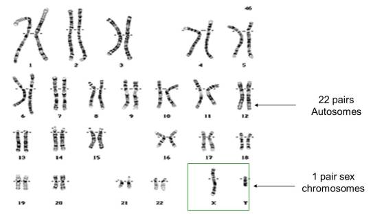
As you can see from the picture above, the 23rd pair of chromosomes in humans are called the sex chromosomes. The person whose chromosomes are shown above is male because he has one X and one Y chromosome in his 23rd pair. If we looked at a picture of a human female set of chromosomes, pairs 1 to 22 would be exactly as above, but the 23rd pair would be different. There would be two large X chromosomes rather than one large X and one tiny Y chromosome as shown above.
So a human female has XX as her 23rd pair of chromosomes, a human male has XY as his 23rd pair.
Gametes (Sperm and Egg cells) are made in a process called Meiosis. Remember that meiosis produces daughter cells that are haploid (this means they only have one member of each pair of chromosomes and so half the genetic material)
When a female cell undergoes meiosis in her ovary, the daughter cells produced (egg cells) will contain one of each of the 23 pairs of chromosomes. For the 23rd pair this will always be an X chromosome since both chromosomes in the 23rd pair are X chromosomes.
When a male cell undergoes meiosis in the testis, the daughter cells produced (sperm cells) will contain one of each of the 22 pairs of chromosomes exactly as above. But the 23 pair are different to each other and so half the sperm cells will contain an X chromosome as the 23rd chromosome and half the sperm cells will contain a Y chromosome as the 23rd chromosome.

If you understand the picture above, you understand sex determination in humans. You also need to be able to draw a genetic diagram to show this.
Phenotype: Mum Dad
23rd pair: XX XY
Gametes: X ½X ½Y
Fertilisation:

Offspring 23rd pair of chromosomes: ½ XX and ½ XY
Offspring phenotypes: ½ female and ½ male
Protein Synthesis (part 3): Grade 9 Understanding for IGCSE Biology 3.18B
This is my final post on protein synthesis, you may be relieved to know…. It is a complicated topic and there is lots to understand but remember it is only one specification point out of several hundred in the IGCSE specification…… So don’t worry too much if you find this hard to grasp and don’t spend a disproportionate amount of time revising it. If you are fascinated by how genetic information is encoded in DNA and how genes work at a molecular level, then you have to choose Biology as a subject to study at A level!
I want to end with two final questions, both of which are essential to address if you are to acquire the grade 9 understanding you need.
What is meant by complementary base pairing and why is it so important?
Let’s go back to the structure of a DNA molecule.

In the middle of the molecule there are pairs of bases. There are four possible bases in DNA – Adenine, Thymine, Cytosine and Guanine – but they are often represented by their letters A,T,C and G.
Bases pair up in a totally predictable way across the double stranded DNA molecule:
A always pairs with T, C always pairs with G. Why is this? Well you can see from the diagram above that A and T are held together by two weak bonds called Hydrogen (H) bonds, whereas C and G are held together by 3 H bonds. This means that this is the only way they can pair up in a stable way.
These pairs of bases (A=T and C≡G) are called complementary base pairs because they always match up in a predictable way.
You can also get complementary base pairing between RNA bases. (Remember that in RNA, there is never any thymine but it is replaced with a different base called uracil) So A=U and C≡G are the complementary base pairs between two RNA molecules.
Complementary base pairing between DNA and RNA bases is essential in transcription but you do not need to know the details at this stage.
We will come back to complementary base pairing in a moment…..
How does the ribosome ensure that the correct amino acids are joined together to make the protein during translation?
To understand this, you need to understand the role that transfer RNA (or tRNA for short) plays in protein synthesis. tRNA molecules are found in the cytoplasm and have an interesting structure. At one end, they have an important triplet of bases called the anticodon. At the other end, there is a place in the molecule where an amino acid can be added.

The key idea is this: the tRNA molecules are loaded up by attaching an amino acid by a group of enzymes found in the cytoplasm. But these enzymes ensure that each amino acid is only attached to tRNA molecules with the correct anticodon.
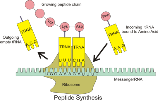
Transfer RNA molecules are so called because they are going to transfer (or carry) the amino acids into the ribosome. You do not need to know the details of protein synthesis but the main idea is that there is complementary base pairing between the anticodon on the tRNA and the codon on the mRNA.
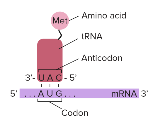
This complementary base pairing between codon and anticodon when combined with the structure of the ribosome means that the amino acids will be joined together in the correct order to make the protein.
Please do not worry too much about the details of all this. I am sure that the examiners will not expect you to know the details of protein synthesis. But you should understand what an an anticodon is and the role of tRNA in the overall process.
anticodon: “a triplet of adjacent bases found on a transfer RNA molecule”
tRNA: “these molecules are found in the cytoplasm of cells and carry the correct amino acid into the ribosome. There is complementary base pairing between the codon on the mRNA and the anticodon on the tRNA as shown in the diagram above”
Protein Synthesis (part 2): Grade 9 Understanding for IGCSE Biology 3.18B
In the last post, I asked you to think of a good “2 mark” explanation of some important terms to do with protein synthesis. Here is my (GCSE level) answer…..
Gene: “a section of a DNA molecule that codes for the production of a single protein”
Ribosome: “a small structure found in the cytoplasm of cells where proteins are made by joining amino acids together into a long chain”
Transcription: “the process occurring in the nucleus in which a double-stranded DNA molecule is used to make a single-stranded molecule of messenger RNA”
Messenger RNA: ” a small single stranded molecule that is made in transcription and can carry the genetic information out of the nucleus to the ribosome”
Translation: “the second stage of protein synthesis that occurs in the cytoplasm in a ribosome in which amino acids are joined together in the correct order to make the protein”
Codon: “a triplet of adjacent bases in an mRNA molecule that codes for a single amino acid”
Finally I want to answer three important questions, one in this post, two in the next….
Why are codons three bases long?
Well, the answer here is a simple bit of Maths. If you remember, there are 20 possible amino acids that can be joined together in any order and in any number to make a protein. In DNA/RNA there are just four bases. So in order to code for 20 amino acids, how many “words” do you need? Well you need at least 20…..
In DNA/RNA you only have 4 “letters” available to make these words. (The letters are the bases and the words are the codons.)
- If the words were one letter long, there are only 4 words. (43 for the mathematicians): A,T,C,G
- If the words were two letters long, there are only 16 possible words (42 for the mathematicians): AA, AT, AC, AG, CA, CT, CC, CG, GA, GT, GC, GG, TA, TT, TC, TG
- If the words were three letters long, there are 64 possible words (43 for the mathematicians) AAA, AAT, AAC, AAG, ACA, ACT, ACC, ACG etc. etc.
So three bases in a codon is a minimum number needed to code for 20 different amino acids. But then this raises a question, doesn’t it? If there are 64 possible words and only 20 words needed, then what happens to all the extra, unnecessary codons? The answer is that there are lots of synonyms in the language of DNA/RNA. (Synonyms are two different words that have the same meaning)
Check out this picture of the genetic code (written in the language of RNA) and notice all the lovely synonyms!

Phe, Leu, Ser, Tyr, Cys and the others are names of amino acids
Notice that almost all amino acids have more than one codon that codes for them. For example, the amino acid Thr (threonine) is coded for by ACU, ACC, ACA and ACG in mRNA.
The three codons marked STOP are used as signals for the ribosome to stop the process of translation (but you don’t need to worry about that unless you wisely choose to continue your studies in Biology at A level!)
This is complicated stuff so please feel free to ask me questions about this using the “leave a comment” box below. You won’t get an instantaneous response but I try to check my blog every couple of days.
RNA: Grade 9 Understanding for IGCSE Biology 3.17B
You need to understand the structure of the molecule DNA before you read this post.
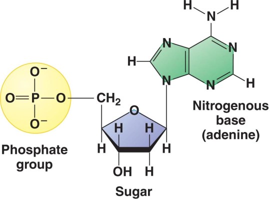
DNA stands for deoxyribonucleic acid and is the chemical that makes up the genetic information in all living organisms on earth. DNA is a double-stranded molecule in which each strand is made of a polymer of simple molecules called nucleotides. There are four nucleotides in DNA, with each nucleotide differing in the base present in the molecule. Adenine, Thymine, Cytosine and Thymine are the four bases found in the nucleotides in DNA. Every nucleotide in DNA contains the same sugar, deoxyribose and a phosphate group as shown in the diagram below.
But DNA is not the only nucleic acid found in cells. All living cells also contain a similar molecule RNA that serves a whole variety of different functions. It is not the main genetic material in any cell but is essential in allowing the information contained in a DNA molecule to be expressed as a protein (see post on protein synthesis to come)
RNA stands for Ribonucleic Acid and is also a polymer of nucleotides. But whereas DNA is always a double-stranded molecule, RNA is always single-stranded (although certain forms of RNA can fold back on themselves at points so they can appear double stranded). The sugar in every RNA nucleotide is ribose (as opposed to deoxyribose in DNA).

There are also four different bases found in RNA nucleotides. Three are identical to the bases found in DNA (Adenine, Cytosine and Guanine) but there is no Thymine in RNA. RNA can contain nucleotides with the similar base Uracil in its place.

So in summary:
- RNA is single stranded whereas DNA is double stranded
- RNA contains the sugar ribose in every nucleotide whereas DNA contains deoxyribose
- DNA contains 4 bases (ATCG) whereas RNA contains A,U,C and G (thymine is replaced by uracil)
Protein Synthesis (part 1): Grade 9 Understanding for IGCSE Biology 3.18B
This is by far the most difficult concept for you to understand in the new GCSE specifications. In fact, it was only ever taught to A level students until last year (and to be honest I would much prefer it that way!). But that is no consolation to you poor folk who are going to get tested on it in your IGCSE and GCSE exams…
I am going to keep this as simple as I possibly can but am not going to dumb it down…. My blog is aimed for students who are ambitious to develop Grade 9 understanding in Biology (this topic is not tested at all in our Double Award Science course) so I want to explain it to you at the level you need. But you will need to read this carefully, take your time and you might need to break it down into small sections to build the understanding you need.
Can I suggest that first of all you read this post from my blog about DNA and how it works?
What is a gene?
A gene is a section of DNA that codes for a single protein. How does this code work? Well the short answer is that the sequence (order) of the bases as you read along the DNA molecule is a code for the sequence of amino acids that are joined together to make the protein.
Remember that there are 4 different bases in DNA: Adenine (A), Thymine (T), Cytosine (C) and Guanine (G). So a sequence of bases on a piece of DNA might look like this:
GCCTATAAATGGCAGGCATTAGCTCTAGGAAATCTAGGGACTTTACA
Protein Synthesis
Proteins are made by joining small molecules called amino acids together. This process is called protein synthesis and happens in small structures in the cytoplasm of all cells called ribosomes. But for all eukaryote cells, this poses a big geographical problem.
The “information” in the gene is stored in a sequence of bases in a DNA molecule and is found in the nucleus. DNA never leaves the nucleus because it is too important a molecule to be allowed into the reactive and unpredictable environment in the cytoplasm. But there are no ribosomes in the nucleus and these are the structures in which proteins are actually made. So a temporary intermediate molecule is needed to carry the “information” from the gene in the nucleus out into the cytoplasm where the ribosomes are found. So the process of making a protein therefore has to exist as a two stage process. The first stage is making the temporary intermediate molecule using the sequence of bases in the gene. Then there is a second stage that happens in the cytoplasm in the ribosome and this involves joining the amino acids together to make the actual protein.
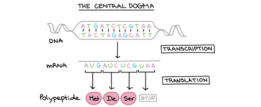
This idea was called the “central dogma of molecular biology” by Watson and Crick in their famous paper on the structure of DNA.

Transcription and Translation
There is quite a lot of jargon in this topic.
Transcription is the name for the process that happens in the nucleus in which a temporary intermediate molecule is made. This temporary “information-containing” molecule is a form of RNA called messenger RNA (or mRNA for short). The mRNA travels out of the nucleus to a ribosome which is found in the cytoplasm. Here a process called Translation occurs in which the the amino acids are joined together in the correct order to make the protein.
Look at this second diagram of the central dogma above. It shows a double-stranded DNA molecule at the top with pairs of bases (either A-T or C-G) joined by hydrogen bonds. The “information” in the molecule is found in the sequence of bases: on the top strand of the DNA this sequence is ATGATCTCGTAA.
You can see that transcription results in the formation of a molecule of mRNA. (Remember that RNA is always a single stranded molecule and contains the base Uracil in place of the base Thymine)
So can you see that the sequence of bases in the mRNA is almost identical to the DNA strand above, but with the base T replaced by the base U.
mRNA sequence: AUGAUCUCGUAA
This diagram shows us one final thing about how protein synthesis works. Look now at the small section of protein (polypeptide) that is produced in translation. You can see that this section of protein is made of three amino acids joined together: methionine (Met), attached to isoleucine (Ile) attached to serine (Ser)
Each amino acid is coded for by a group of 3 adjacent bases on the mRNA molecule. These triplets of bases are called Codons.
- AUG is a codon that codes for the amino acid Methionine
- AUC is a codon that codes for the amino acid Isoleucine
- UCG is a codon that codes for the amino acid Serine
(UAA is called a stop codon as it ends the translation process at the ribosome)
A codon is a triplet of adjacent bases on a mRNA molecule. Each codon codes for a single amino acid that will be joined together to make the protein.
Check your understanding:
Can you explain the meaning of the following terms? Write a 2 mark explanation of what each word means.
- Gene
- Ribosome
- Transcription
- Messenger RNA
- Translation
- Codon
I will put the answers into the next post called Protein Synthesis (part 2) which I promise I will write tomorrow…… That’s enough for now.
Menstrual Cycle video
This is a great summary video on the hormones of the menstrual cycle. IGCSE students should not worry about the role of GnRH from the hypothalamus, but should instead focus on the hormones FSH, LH, oestrogen and progesterone.

Differences between Sexual and Asexual Reproduction: Grade 9 Understanding for IGCSE Biology 3.1 3.2
All organisms have the potential to reproduce. Reproduction is one of the 7 characteristics of life (8 if you study EdExcel IGCSE Biology…..) and of course it means the ability to produce new individuals. But over the 4 billion or so years life has been around on the planet, evolution has developed a myriad ways of producing new individuals. So as biologists are simple folk (well all the ones I work with are….), it makes sense to group different reproductive strategies together to make it easier to understand.
The major distinction between different ways of reproducing is to divide them into asexual and sexual reproduction. This works fairly well, although it seems to be a subject many GCSE students don’t understand too well. So here goes….
The first big idea to dispel is that the number of parents involved determines whether reproduction is sexual or asexual. Too often I am told that asexual involves one parent, sexual two. But although this works in most cases, sexual reproduction can happen with only one parent, for example in flowering plants that self-pollinate. So we need a different way of deciding whether reproduction is sexual or asexual.
And in fact a clear distinction does exist and it is to do with genetics. If the offspring produced are genetically identical to the parent (i.e. a clone) then it is an asexual form of reproduction. If the offspring produced are genetically different to the parents, then it is sexual.
Often sexual reproduction involves the process of fertilisation. This allows two parents to each contribute half their genetic material to their offspring thus generating individuals with new and unique genetic make ups. These specialised cells that contain half the genetic material are called gametes and as you all know, they are made by a special type of cell division called meiosis. Meiosis is vital for sexual reproduction as it produces cells that are haploid (one member of each pair of chromosomes) and all genetically unique.
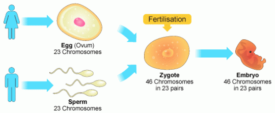
So this diagram shows two humans each producing gametes by meiosis. The parents on the left will have 23 pairs of chromosomes and so each haploid gamete will have 23 individual chromosomes. Fertilisation restores the diploid number. Mitosis is then used to turn this single cell, the zygote into a multicellular embryo and then indeed into a new individual. (You will remember mitosis is a type of cell division that always produces genetically identical daughter cells)
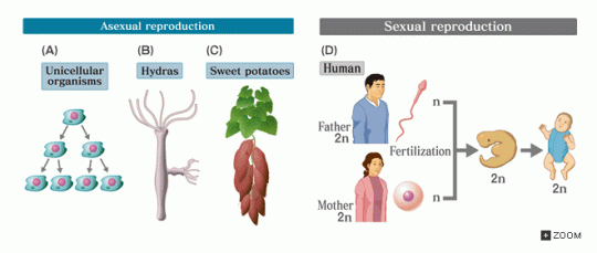
The examples of asexual reproduction on the left all involve only this second type of cell division, mitosis. There are no gametes, no fertilisation and hence no genetic variation. The simplest type of asexual reproduction is shown as (A) and this is called binary fission. A single-celled organism can divide in two to produce two genetically identical daughter cells. Hydra (B) are a simple type of animal and they reproduce by budding. A new individual just grows off the side and when it is big enough, it drops off….. And many plants can reproduce asexually using a technique called vegetative propagation. The sweet potato plant in (C) can produce several offspring plants from each potato but as they are all clones of each other, this is definitely asexual reproduction.
Students do get confused with this topic so please ask me a question using the comment feature below the post. Keep revising hard!

Variation: Grade 9 Understanding for IGCSE Biology 3.33
Variation within a population is a critical and required component for natural selection. If you have understood all the work on DNA, chromosomes and Mendelian genetics, you should now have a good understanding of where the genetic causes of this variation comes from. But remember that variation can also be caused by the environment. Indeed all variations in reality come from an interplay between genetic and environmental factors. Genes by themselves cannot cause variation as without the environment of a cell to produce the protein, genes alone cannot affect the phenotype.
Genetic causes of variation
Sexual reproduction is the key to genetic variation in a population. Meiosis produces gametes that are haploid and genetically different from each other. The gametes may have a different combination of randomly “shuffled” chromosomes. Crossing over in meiosis also allows alleles that would not otherwise be combined in a gamete. So there are massive genetic differences between one gamete and the next. And then there is random fertilisation so that any one male gamete is equally likely to fuse with any female gamete. Random fertilisation (of gametes that meiosis has made genetically different to each other) is the key to genetic variation in a population.
A final cause of genetic variation has nothing to do with sexual reproduction and is mutation. DNA replication is not 100% accurate – the enzymes in eukaryotes make one error every billion base-pairs. These mutations are random and can lead to new alleles appearing in a population. Chromosomal mutations can also occur where chromosomes do not separate properly in meiosis or parts of a chromosome break off and re-join somewhere else….
Environmental causes of variation
Some of the differences seen in populations are due not to differences in genes but due to the differing environments in which an organism lives. Peas which have inherited two copies of the T allele (for tallness) will never grow tall unless they are planted in well-watered soil and given access to sunlight. You would never be a school teacher as a career without understanding that the environment a brain develops in can affect a person’s outcomes. Environment is as important as genes in many variations in the human population, in particular to do with health and disease. This is why so much emphasis for health for example is placed on promoting balanced diets, altering smoking habits, and moderating alcohol consumption.
Genes and Environment always interact to determine Phenotype
Don’t allow yourself to fall into the lazy thinking of the “nature-nurture” debate. It is lazy thinking because the debate is a nonsense. Nothing is either determined by your genes or your environment – it is always both. So when you read of a ‘gene for obesity’ or a ‘gene for domestic violence’ treat with extreme caution (and switch newspapers……) If you read that playing violent computer games causes violent behaviour in humans, treat with caution. None of these variations in a population will be just due to genes, none will be just do to the environment. It will always be some complex interaction between the two.

Cell Division part 3: Grade 9 Understanding of Meiosis for IGCSE 3.30
In sexually reproducing organisms two types of cell division are needed. One is for the processes of growth, repair and asexual reproduction and it is called mitosis. Mitosis produces daughter cells that are diploid and genetically identical to the parent cell.
But when the organism wants to make gametes a different mechanism is needed. Gametes are not diploid like all the other body cells, but instead they only have one member of each homologous pair of chromosomes. In order to make a haploid daughter cell, a second type of cell division, meiosis, is needed.

You can see in the diagram above some of the key differences between mitosis and meiosis. Both start with diploid cells (2n) but whereas mitosis involves one round of division and produces two identical diploid daughter cells, meiosis is different. Meiosis has two rounds of division, called Meiosis I and Meiosis II. This results in four daughter cells and you can see that they are all haploid (n) cells. These cells develop into gametes (sperm and egg cells in humans) and so when they fuse together in fertilisation, the diploid number is restored.

Gametes are all genetically different to each other
Meiosis does not just produce haploid daughter cells. It also introduces genetic variation into the daughter cells so each is genetically unique. This means that random fertilisation will produce offspring that are all genetically different to either parent. How does this genetic variation in gametes come about?
Well to answer that, you need to understand a little bit more about how the chromosomes behave during meiosis. I am not going to talk through all the various stages of meiosis (life is too short and you can read the diagram below) but there is a key event that happens in meiosis that never happens in mitosis…..
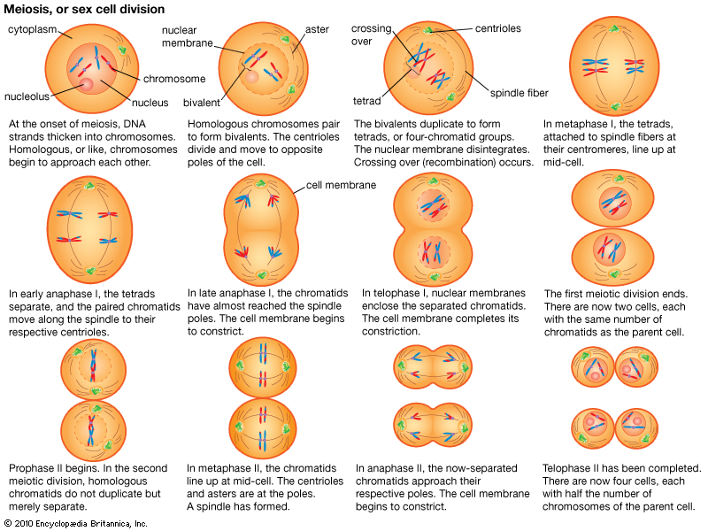
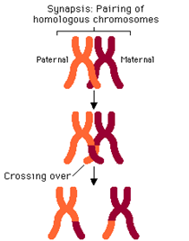
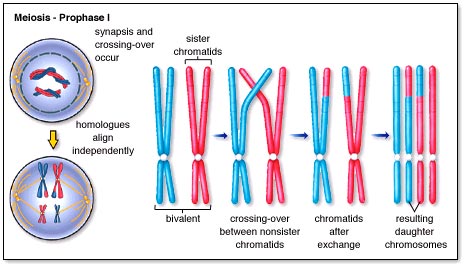
It happens in prophase of the first meiotic division and is called synapsis. As the nuclear membrane is degenerating, the two members of a homologous pair of chromosomes line up alongside each other to form a structure called a bivalent (or tetrad)
In the first meiotic division, the two members of the homologous pair are pulled apart and separated. Because these bivalents attach and assort independently of each other, this means that this random assortment can produce many different gametes. A human cell with 23 pairs of chromosomes can produce 2^23 possible gametes just by random assortment.
But there is a second process called crossing over that happens during prophase 1 when the bivalents are formed. As you can see in the diagrams, small sections of chromatid can be swapped between the chromatids of one chromosome and with its homologous partner. This ensures that when the individual chromatids are separated in meiosis 2, each is different to each other. This multiplies up the genetic variation by several orders of magnitude. (see diagram below)
Now that is more detailed than you will need in an iGCSE exam, but it is good to understand where the genetic variation in gametes comes from. Let’s finish with something more simple – the differences between mitosis and meiosis.
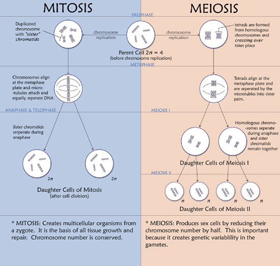
One final point: please learn the spellings of these two types of cell division. Spelling is only penalised in exams when the meaning is lost and any intermediate spelling (e.g. meitosis or miosis) has no meaning! So if you are one of those people who finds spelling difficult, find a way of learning mitosis (produces identical diploid daughter cells and is used in growth) compared to meiosis (produces genetically different haploid cells and is used to make gametes)
