
Tagged: insulin
The Human Alimentary canal: Grade 9 Understanding for IGCSE Biology 2.27
A human body is in many ways rather like a packet of polo mints. These are famous mints in the UK for having a hole in the middle. Our body is divided into segments (rather like the packet of polos) and we have a tube that runs through the middle of us. This tube is called the Alimentary Canal (or Gut) and it’s function in the body is the digestion and absorption of food molecules (see later post on “Stages of Processing Food”)

The Alimentary Canal is divided into specialised regions, each with its own particular range of functions to do with the processing of food. You are required to understand a little about some of these organs and their functions.

The first thing is to make sure you can label a diagram of the human digestive system such as the one shown above. Check that you could accurately identify the following structures:
mouth, tongue, teeth, salivary glands, oesophagus, stomach, liver, gall bladder, bile duct, pancreas, pancreatic duct, duodenum, ileum, colon, appendix, rectum, anus
Here is a good diagram to use to check your labelling of the human digestive system

Functions
1 Mouth
The mouth is actually the name for the opening at the top of the alimentary canal rather than the chamber behind. If you want to be really precise, you should call this chamber containing the tongue and teeth by its proper name, the buccal cavity. The mouth is the opening that allows an animal to ingest food. In the buccal cavity, the teeth can chop up the food into smaller pieces and the tongue can move the food into a ball (bolus) for swallowing. The food is tasted in the buccal cavity and there are many chemoreceptors on the tongue and in the nasal cavity that perform this function. There are three sets of salivary glands around the buccal cavity and these secrete a watery liquid, saliva to mix with the ingested food. Saliva is alkaline to help protect the tooth enamel from acidic decay by bacteria but also contains a digestive enzyme, salivary amylase that begins the process of digestion of starch in the mouth. Salivary amylase catalyses the hydrolysis reaction in which starch, a polysaccharide is digested into the disaccharide maltose.
2 Oesophagus
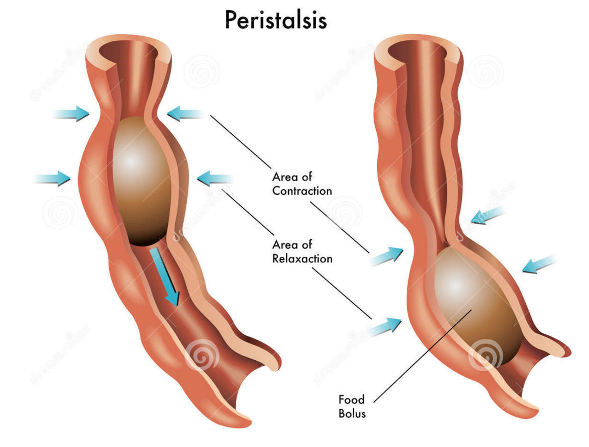
The oesophagus is the tube that carries the ball of food from the back of the throat through the thorax and down into the stomach. The Alimentary canal has layers of muscle in its wall throughout its entire length. These layers of smooth muscle can contract and relax in an antagonistic fashion to push the bolus along the tube. There are two main types of smooth muscle in the wall of the Alimentary canal – circular fibres are arranged around the circumference of the tube and longitudinal fibres are arranged along the length of the tube. These waves of alternate contraction and relaxation are called peristalsis.

3 Stomach
The stomach is a muscular storage organ that keeps food in it for around 3-4 hours before squirting it out in small amounts into the duodenum. The muscle layers in the stomach wall churn the food and mix it with the secretions from the stomach lining. These secretions are called gastric juice and contain a mixture of hydrochloric acid, mucus and a digestive enzyme pepsin. The acid makes the gastric juice overall very acidic, around pH 1.5. This acidity forms part of the non-specific defences of the body against bacteria as the extreme pH kills almost all bacteria in the food. The mucus is important as it protects the cells lining the stomach from the acidity. Pepsin is a digestive enzyme that starts the digestion of protein. It catalyses a hydrolysis reaction in which proteins are broken down into smaller molecules called polypeptides. Pepsin is an unusual enzyme in that it has an optimum pH of around 1.5.
4 Small Intestine
I will write a whole post on the small intestine later this week as there is plenty for you to understand about this part of the alimentary canal. All I will say here is that it is divided into the duodenum which is where almost all the digestion reactions take place and the ileum which is adapted for efficient absorption of the products of digestion into the blood. (see post later in the week if you want to find out more….)
5 Large Intestine
The majority of the large intestine is made up of an organ called the colon. The colon has a variety of functions. It is where water from all the various secretions is reabsorbed back into the blood, thus producing a solid waste called faeces. (Water that you drink tends to be absorbed through the stomach lining much earlier in the alimentary canal) There are also a few mineral salts and vitamins absorbed into the bloodstream in the colon. The colon is also home to a varied population of bacteria, the so called gut flora. Faeces is stored in the final part of the large intestine that is called the rectum.
6 Pancreas
The pancreas is not part of the alimentary canal (although I am not sure the person who wrote the specification appreciated that….) It is an example of what is called an accessory organ for the digestive system. The pancreas is a really interesting organ as it contains different cell types that carry out two completely separate functions. The majority of the cells in the pancreas secrete a whole load of digestive enzymes into an alkaline secretion called pancreatic juice. There is a tube called the pancreatic duct that carries the pancreatic juice and empties it into the duodenum where it can mix with the acidic chyme coming out of the stomach.
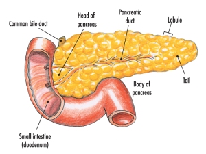
There are small clusters of a different kind of cell found in the pancreas. These are the islets of Langerhans that secrete the hormones insulin and glucagon into the bloodstream. These two pancreatic hormones together regulate the blood glucose concentration.
Hormones: Grade 9 Understanding for IGCSE Biology 2.94 2.95B
Hormones are defined as “chemicals produced in endocrine glands that are secreted into the bloodstream and cause an effect on target tissues elsewhere in the body”. They play a wide variety of roles in the healthy functioning and development of the body.

The iGCSE specification only really mentions a small number of hormones so these are the ones I will focus on in this post.
ADH (anti-diuretic hormone) (Separate Biologists only – not Combined Science)
ADH is secreted into the blood by an endocrine gland at the base of the brain called the Pituitary Gland. The stimulus for the release of ADH into the blood comes from the hypothalamus (a region of brain right next to the pituitary gland) when it detects that the blood plasma is becoming too concentrated. This might be caused by the body becoming dehydrated due to sweating. ADH travels round the body in the blood until it reaches its target tissue which are the cells that line the collecting ducts in the nephrons in the kidney. ADH increases the permeability of the connecting duct walls to water, thus meaning more water is reabsorbed by osmosis from the urine in the collecting duct and back into the blood. This results in a small volume of concentrated urine being produced.
Adrenaline
Adrenaline is secreted into the blood by the adrenal glands in situations of danger or stress.. The adrenals are found just above the two kidneys on the back of the body wall. Adrenaline secretion is controlled by nerve cells that come from the central nervous system. Adrenaline is often described as the “fight or flight” hormone as its effects are to prepare the body to defend itself or run away from danger. There are receptors for adrenaline in many target tissues in the body but some of the most significant effects of adrenaline are:
- affects the pacemaker cells in the heart causing an increase in heart rate
- shifts the pattern of blood flow into muscles, skin and away from the intestines and other internal organs
- decreases peristalsis in the gut
- causes pupils to dilate in the eye
- increases breathing rate in the lungs
- promotes the passing of urine from the bladder
Insulin
Insulin is a hormone made in the islets of Langerhans in the pancreas. It plays a vital role in the homeostatic control of the blood sugar concentration. The pancreas will secrete insulin into the blood when the blood glucose concentration gets too high. There are many cells in the body with insulin receptors but the main target tissue for insulin is the liver.
Insulin causes the liver (and muscle) cells to take glucose out of the blood and convert it into the storage polysaccharide glycogen. This results in a lowering of the blood glucose concentration: a good example of the importance of the principle of negative feedback in homeostasis
Testosterone
Testosterone is a steroid hormone made by cells in the testes of males. It is the main hormone of puberty in males resulting in the growth of the reproductive organs at puberty as well as the secondary sexual characteristics (pitch of voice lowering, muscle growth stimulated, body hair grows etc.)
Oestrogen
Oestrogen is a steroid hormone made by the cells in the ovary that surround the developing egg cell in the first half of the menstrual cycle. In puberty it causes the development of the female secondary sexual characteristics (breast growth, change in body shape, pubic hair etc.) but in the menstrual cycle, oestrogen has a variety of important effects. It stimulates the rebuilding of the uterine endometrium (or lining) to prepare the uterus for the implantation of an embryo. Oestrogen also affects the pituitary gland and can cause the spike in LH concentrations that trigger ovulation on day 14 of the cycle.
Progesterone
Progesterone is also made in the ovary but at a different time in the menstrual cycle. It is secreted by cells in the corpus luteum, a structure found from day 14 onwards after the egg has been released in ovulation. Progesterone has two main target tissues: it maintains the thickened lining of the endometrium in the uterus ready for implantation. Progesterone also causes the pituitary gland to stop secreting the hormones FSH and LH so a new cycle is never started. It is for this reason that progesterone can be used in women as a contraceptive pill.
FSH (Follicle-Stimulating Hormone (Separate Biologists only – not Combined Science)
FSH is a hormone released by the pituitary gland underneath the brain. The target tissues for FSH are in the testis (males) and ovaries (females). In males FSH plays a role in the growth of the testes allowing sperm production to start. In females, FSH is the hormone released at the start of the menstrual cycle that causes one of the immature egg cells in an ovary to grow, develop and so become surrounded by follicle cells prior to ovulation.
LH (Luteinising Hormone) (Separate Biologists only – not Combined Science)
LH is a second reproductive hormone released by the pituitary gland into the bloodstream. In males, it stimulates the production of testosterone in the testes. In females, it is released only on days 13 and 14 of the menstrual cycle and it is the hormone that triggers ovulation.
The Human Alimentary canal: Grade 9 Understanding for IGCSE Biology 2.27
A human body is in many ways rather like a packet of polo mints. These are famous mints in the UK for having a hole in the middle. Our body is divided into segments (rather like the packet of polos) and we have a tube that runs through the middle of us. This tube is called the Alimentary Canal (or Gut) and it’s function in the body is the digestion and absorption of food molecules (see later post on “Stages of Processing Food”)
The Alimentary Canal is divided into specialised regions, each with its own particular range of functions to do with the processing of food. You are required to understand a little about some of these organs and their functions.
The first thing is to make sure you can label a diagram of the human digestive system such as the one shown above. Check that you could accurately identify the following structures:
mouth, tongue, teeth, salivary glands, oesophagus, stomach, liver, gall bladder, bile duct, pancreas, pancreatic duct, duodenum, ileum, colon, appendix, rectum, anus
Here is a good diagram to use to check your labelling of the human digestive system
Functions
1 Mouth
The mouth is actually the name for the opening at the top of the alimentary canal rather than the chamber behind. If you want to be really precise, you should call this chamber containing the tongue and teeth by its proper name, the buccal cavity. The mouth is the opening that allows an animal to ingest food. In the buccal cavity, the teeth can chop up the food into smaller pieces and the tongue can move the food into a ball (bolus) for swallowing. The food is tasted in the buccal cavity and there are many chemoreceptors on the tongue and in the nasal cavity that perform this function. There are three sets of salivary glands around the buccal cavity and these secrete a watery liquid, saliva to mix with the ingested food. Saliva is alkaline to help protect the tooth enamel from acidic decay by bacteria but also contains a digestive enzyme, salivary amylase that begins the process of digestion of starch in the mouth. Salivary amylase catalyses the hydrolysis reaction in which starch, a polysaccharide is digested into the disaccharide maltose.
2 Oesophagus
The oesophagus is the tube that carries the ball of food from the back of the throat through the thorax and down into the stomach. The Alimentary canal has layers of muscle in its wall throughout its entire length. These layers of smooth muscle can contract and relax in an antagonistic fashion to push the bolus along the tube. There are two main types of smooth muscle in the wall of the Alimentary canal – circular fibres are arranged around the circumference of the tube and longitudinal fibres are arranged along the length of the tube. These waves of alternate contraction and relaxation are called peristalsis.
3 Stomach
The stomach is a muscular storage organ that keeps food in it for around 3-4 hours before squirting it out in small amounts into the duodenum. The muscle layers in the stomach wall churn the food and mix it with the secretions from the stomach lining. These secretions are called gastric juice and contain a mixture of hydrochloric acid, mucus and a digestive enzyme pepsin. The acid makes the gastric juice overall very acidic, around pH 1.5. This acidity forms part of the non-specific defences of the body against bacteria as the extreme pH kills almost all bacteria in the food. The mucus is important as it protects the cells lining the stomach from the acidity. Pepsin is a digestive enzyme that starts the digestion of protein. It catalyses a hydrolysis reaction in which proteins are broken down into smaller molecules called polypeptides. Pepsin is an unusual enzyme in that it has an optimum pH of around 1.5.
4 Small Intestine
I will write a whole post on the small intestine later this week as there is plenty for you to understand about this part of the alimentary canal. All I will say here is that it is divided into the duodenum which is where almost all the digestion reactions take place and the ileum which is adapted for efficient absorption of the products of digestion into the blood. (see post later in the week if you want to find out more….)
5 Large Intestine
The majority of the large intestine is made up of an organ called the colon. The colon has a variety of functions. It is where water from all the various secretions is reabsorbed back into the blood, thus producing a solid waste called faeces. (Water that you drink tends to be absorbed through the stomach lining much earlier in the alimentary canal) There are also a few mineral salts and vitamins absorbed into the bloodstream in the colon. The colon is also home to a varied population of bacteria, the so called gut flora. Faeces is stored in the final part of the large intestine that is called the rectum.
6 Pancreas
The pancreas is not part of the alimentary canal (although I am not sure the person who wrote the specification appreciated that….) It is an example of what is called an accessory organ for the digestive system. The pancreas is a really interesting organ as it contains different cell types that carry out two completely separate functions. The majority of the cells in the pancreas secrete a whole load of digestive enzymes into an alkaline secretion called pancreatic juice. There is a tube called the pancreatic duct that carries the pancreatic juice and empties it into the duodenum where it can mix with the acidic chyme coming out of the stomach.
There are small clusters of a different kind of cell found in the pancreas. These are the islets of Langerhans that secrete the hormones insulin and glucagon into the bloodstream. These two pancreatic hormones together regulate the blood glucose concentration.

Recombinant DNA: Grade 9 Understanding for IGCSE Biology 5.12 5.13 5.14
In the last post on this topic, I explained about the two types of enzymes needed for the genetic modification of organisms:
Restriction enzymes that can cut up DNA molecules at specific target sequences, often resulting in fragments with sticky ends
DNA ligase that joins together fragments to form a single DNA molecule
The EdExcel iGCSE syllabus uses the example of the genetic modification of bacteria to produce human insulin. Human insulin is a hormone that helps regulate the concentration of glucose in the blood. It is made in the pancreas when the blood glucose concentration gets too high and causes liver cells to take up glucose from the blood and convert it to the storage molecule glycogen. Patients with type I diabetes cannot make their own insulin and so need to inject it several times a day after meals to ensure they maintain a constant healthy concentration of glucose in their blood.
Bacteria can be genetically modified so that they produce human insulin. These transgenic bacteria can be cultured in a fermenter and the insulin produced can be extracted, purified and sold.
How do you get hold of the human insulin gene?
Well the honest answer is that there are a variety of ways of achieving this. It can now be synthesised artificially as we know the exact base sequence of the gene but it can also be cut out of a human DNA library using a restriction enzyme. There are other ways too but for the sake of brevity (and sanity) I am not going to go into them here. [If you are really interested in this, find out how reverse transcription of messenger RNA from cells in the pancreas can allow you to build the insulin gene.]
How do you get the human insulin gene into a bacterium?
Remember that bacterial cells are fundamentally different to animal and plant cells. One difference is that bacterial cells have no nucleus and their DNA is in the form of a ring that floats in the cytoplasm. Many bacteria also have plasmids which are small additional rings of DNA and these provide a way for getting a new gene into a bacterium.

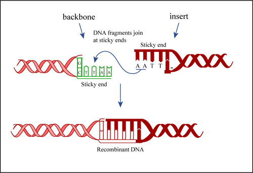
Bacteria exchange plasmids in a process called conjugation and so it is fairly easy to get the plasmid out of the bacterium. If the plasmid is cut open using the same restriction enzyme as was used to cut out the human insulin gene, the sticky ends will match up and so DNA ligase will join the two pieces of DNA together to make a recombinant plasmid. The diagram below shows the process for human growth hormone but it would be exactly the same for the example we are looking at.
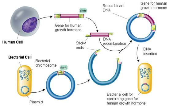
If the recombinant plasmids are inserted into bacteria, the bacteria will read the human insulin gene and so produce the protein insulin.

How do you grow the transgenic bacteria on an industrial scale?
The bacteria that have taken up the recombinant plasmid are grown in a fermenter. This is a large stainless steel vat (easy to clean and sterilise) that often has several design features conserved between different varieties:

The fermenter usually has a cooling jacket to carry away excess heat. The jacket often has a cold water input pipe and the warmer water is carried away. There has to be some mechanism for mixing the contents of the fermenter so the diagram above shows paddles attached to a motor. Fermenters also need a sterile input system for getting air, water and nutrients into the fermenter but without introducing foreign bacteria and fungi. Air is needed as the bacteria are aerobic and need oxygen for respiration.

If the bacteria in the fermenter contain the human insulin gene, then they will be able to produce human insulin. This can be extracted, purified and sold to the NHS for treating type I diabetics.

Genetic Modification Grade 9 Understanding for IGCSE Biology 5.12 5.13 5.14 5.16
One of the most complicated areas in the iGCSE course is looking at how organisms can be genetically modified. Remember that humans have been messing around with the genetic composition of many species for thousands of years. Up until recently this has only been using a technique called selective breeding or artificial selection.
Make sure you understand exactly what is meant by the term selective breeding? You probably should be able to explain at least one example in both an animal and a plant species.
In the twentieth century scientists developed a much more precise way of genetically modifying a species. This was due to discoveries about the nature of the genetic code and also the existence of two types of enzyme that make cutting up and then sticking together pieces of DNA. This new technique was called genetic engineering and it has two big advantages over selective breeding. Firstly genes from different species can be recombined to form transgenic organisms. Transgenic is an important term and means an organism that contains DNA from more than one species. This means scientists are not restricted to alleles present in the natural population but can insert genes from any species into any other. Secondly, selective breeding has a big disadvantage in that it can reduce the allelic diversity in a population. If the population becomes more and more similar at a genetic level, this means that inbreeding becomes more of a problem and the population becomes susceptible to damage from changes in the environment.
You need to understand the role of two enzymes in the process of Genetic Engineering: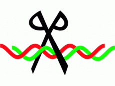
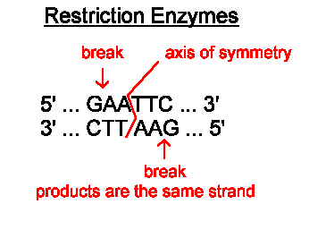
Restriction Enzymes are found in bacteria and have evolved to combat viral infection in the bacterial cell. These enzymes can cut double-stranded DNA at a specific target sequence, often leaving the ends of the DNA with short sections of unpaired bases. These are called sticky ends.
The image below shows the cutting site of a restriction enzyme called EcoR1. You can see the enzyme cuts the DNA anywhere the following sequence is found GAATTC. The DNA molecule is cut after the first G, leaving the two strands with four unpaired bases that make up the two sticky ends.
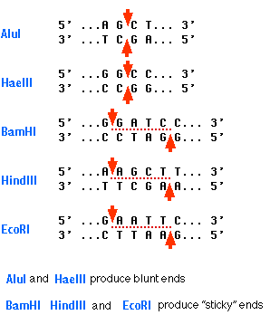
Here are some more restriction enzymes and there target sequences. The bottom three on the list all produce sticky ends.
The second important enzyme for genetic engineering is DNA Ligase. This enzyme catalyses the joining together of the sticky ends of two fragments to produce an intact DNA molecule.
The starting point for understanding the complex topic of genetic modification of organisms is understanding the role of these two enzymes in the process.
Hormones: Grade 9 Understanding for IGCSE Biology 2.94 2.95B
Hormones are defined as “chemicals produced in endocrine glands that are secreted into the bloodstream and cause an effect on target tissues elsewhere in the body”. They play a wide variety of roles in the healthy functioning and development of the body.
The iGCSE specification only really mentions a small number of hormones so these are the ones I will focus on in this post.
ADH (anti-diuretic hormone) (Separate Biologists only – not Combined Science)
ADH is secreted into the blood by an endocrine gland at the base of the brain called the Pituitary Gland. The stimulus for the release of ADH into the blood comes from the hypothalamus (a region of brain right next to the pituitary gland) when it detects that the blood plasma is becoming too concentrated. This might be caused by the body becoming dehydrated due to sweating. ADH travels round the body in the blood until it reaches its target tissue which are the cells that line the collecting ducts in the nephrons in the kidney. ADH increases the permeability of the connecting duct walls to water, thus meaning more water is reabsorbed by osmosis from the urine in the collecting duct and back into the blood. This results in a small volume of concentrated urine being produced.
Adrenaline
Adrenaline is secreted into the blood by the adrenal glands in situations of danger or stress.. The adrenals are found just above the two kidneys on the back of the body wall. Adrenaline secretion is controlled by nerve cells that come from the central nervous system. Adrenaline is often described as the “fight or flight” hormone as its effects are to prepare the body to defend itself or run away from danger. There are receptors for adrenaline in many target tissues in the body but some of the most significant effects of adrenaline are:
- affects the pacemaker cells in the heart causing an increase in heart rate
- shifts the pattern of blood flow into muscles, skin and away from the intestines and other internal organs
- decreases peristalsis in the gut
- causes pupils to dilate in the eye
- increases breathing rate in the lungs
- promotes the passing of urine from the bladder
Insulin
Insulin is a hormone made in the islets of Langerhans in the pancreas. It plays a vital role in the homeostatic control of the blood sugar concentration. The pancreas will secrete insulin into the blood when the blood glucose concentration gets too high. There are many cells in the body with insulin receptors but the main target tissue for insulin is the liver.
Insulin causes the liver (and muscle) cells to take glucose out of the blood and convert it into the storage polysaccharide glycogen. This results in a lowering of the blood glucose concentration: a good example of the importance of the principle of negative feedback in homeostasis
Testosterone
Testosterone is a steroid hormone made by cells in the testes of males. It is the main hormone of puberty in males resulting in the growth of the reproductive organs at puberty as well as the secondary sexual characteristics (pitch of voice lowering, muscle growth stimulated, body hair grows etc.)
Oestrogen
Oestrogen is a steroid hormone made by the cells in the ovary that surround the developing egg cell in the first half of the menstrual cycle. In puberty it causes the development of the female secondary sexual characteristics (breast growth, change in body shape, pubic hair etc.) but in the menstrual cycle, oestrogen has a variety of important effects. It stimulates the rebuilding of the uterine endometrium (or lining) to prepare the uterus for the implantation of an embryo. Oestrogen also affects the pituitary gland and can cause the spike in LH concentrations that trigger ovulation on day 14 of the cycle.
Progesterone
Progesterone is also made in the ovary but at a different time in the menstrual cycle. It is secreted by cells in the corpus luteum, a structure found from day 14 onwards after the egg has been released in ovulation. Progesterone has two main target tissues: it maintains the thickened lining of the endometrium in the uterus ready for implantation. Progesterone also causes the pituitary gland to stop secreting the hormones FSH and LH so a new cycle is never started. It is for this reason that progesterone can be used in women as a contraceptive pill.
FSH (Follicle-Stimulating Hormone (Separate Biologists only – not Combined Science)
FSH is a hormone released by the pituitary gland underneath the brain. The target tissues for FSH are in the testis (males) and ovaries (females). In males FSH plays a role in the growth of the testes allowing sperm production to start. In females, FSH is the hormone released at the start of the menstrual cycle that causes one of the immature egg cells in an ovary to grow, develop and so become surrounded by follicle cells prior to ovulation.
LH (Luteinising Hormone) (Separate Biologists only – not Combined Science)
LH is a second reproductive hormone released by the pituitary gland into the bloodstream. In males, it stimulates the production of testosterone in the testes. In females, it is released only on days 13 and 14 of the menstrual cycle and it is the hormone that triggers ovulation.
