
Tagged: pregnancy
Menstrual Cycle video
This is a great summary video on the hormones of the menstrual cycle. IGCSE students should not worry about the role of GnRH from the hypothalamus, but should instead focus on the hormones FSH, LH, oestrogen and progesterone.
Female Reproductive System: Grade 9 Understanding for IGCSE Biology 3.8
The human male and female reproductive systems are made from the same embryonic cells and are perhaps more similar in structure and function than is first apparent. There are two ovaries protected within the pelvic cavity. The ovary is the site of egg cell production. The egg cell is the female gamete and is haploid – it has only one chromosome from each homologous pair. The ovaries are also endocrine organs that produce the female sex hormones oestrogen and progesterone.
[Indeed differences between the gametes is the essential difference between male and female organisms. Females are always individuals who produce a small number of large, often immobile gametes. You can easily remember this: female – few, fixed, fat. Males are organisms that produce large numbers of small, motile games. Male – many, mini, motile.]
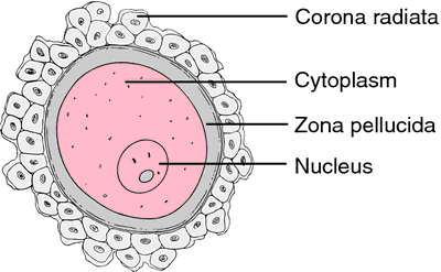
This diagram shows the human egg cell after it has been released from the ovary into the Fallopian tubes (or oviduct). The egg cell is coloured pink in the diagram above (if you are being picky it is not really an egg but a cell called a secondary oocyte but I won’t stress over this now…) The egg cell is surrounded by a thick jelly-like layer called the zona pellucida and then by a whole cluster of mother’s cells from her ovary – the corona radiata. The big idea to remember is that the egg cell is very large compared to sperm cells: it is one of the largest cells in humans with a diameter of about 500 micrometers.
The Fallopian tubes carry the egg down towards the uterus. The lining of the Fallopian tubes is covered in a ciliated epithelium. The cilia waft to generate a current that helps move the egg down towards the uterus. Sperm cells have to swim against this current to reach the egg in the tubes. The Fallopian tube is the usual site for fertilisation to occur.
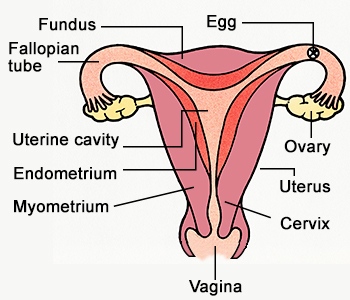
Once fertilisation has occurred, the newly formed zygote divides over and over again by mitosis to form a ball of cells called an embryo. The embryo continues its journey down the Fallopian tube until it reaches the uterus. The uterus (womb) is a muscular organ with a thickened and blood-rich lining called the endometrium. Implantation occurs when the embryo attaches to the endometrium and over time, a placenta forms. The embryo develops into a foetus and remains in the uterus for 9 months.
The cervix is a narrow opening between the uterus and the vagina. It holds the developing foetus in the uterus during pregnancy but dilates (widens) at birth to form part of the birth canal. The vagina is the organ into which sperm are deposited from the man’s penis during sexual intercourse. The lining of the vagina is acidic to protect against bacterial pathogens and the sperm cells released into the vagina quickly start to swim away from the acidity in grooves in the lining. These grooves lead to the cervix and hence into the uterus.

Role of the Amnion – Grade 9 Understanding for IGCSE Biology 3.12
Once the embryo has reached the uterus 7 days after fertilisation, it can implant into the thickened, sticky and blood-rich endometrium. The implanted embryo grows into the uterine lining and starts to surround itself with a collection of membranes. Some of these membranes develop into structures in the placenta, but one the amnion has a different function altogether. The amnion produces a fluid called amniotic fluid that cushions the developing embryo and foetus right through pregnancy and to birth.

The main advantage of having the developing embryo in a sac of amniotic fluid is that it protects the embryo by cushioning against blows to the abdomen. It is also essential for allowing the foetus to move around inside the uterus thus allowing development of the muscular system. The amniotic fluid enters the babies lungs and can promote normal development there. The foetus will swallow amniotic fluid into its stomach and will produce urine into the amniotic fluid as well. Disgusting I know, but that’s babies for you……..
Amniotic fluid contains stem cells. In the future it may be possible to harvest these pluripotent cells and use them to create adult tissues for medical uses.
Role of the Placenta – Grade 9 Understanding for IGCSE Biology 3.11
There are two syllabus points in bold (only tested in paper 2) that refer to embryonic and foetal development. The first asks you to understand the role of the placenta in supplying the developing foetus with nutrients and oxygen and the second concerns the role of amniotic fluid in protecting the developing embryo.
Placenta
The placenta is in many ways a remarkable organ. It contains a mixture of maternal cells from the uterine lining and embryonic cells, but these cells from two genetically different individuals are capable of sticking together to form the placenta. The placenta is only present in the uterus once an embryo has successfully implanted a week or so after fertilisation has happened in the Fallopian Tubes. The placenta is linked to the foetus via the umbilical cord, a structure that contains an umbilical artery and vein carrying foetal blood to and from the placenta.

There is a key idea here that is very important. There is no mixing of maternal and foetal blood in the placenta. This would be disastrous for both mother and baby for a whole variety of reasons. The maternal blood is at a much higher pressure than the foetal blood and if the foetus were connected to the maternal circulatory system directly, its blood vessels would burst. The foetus and mother can have different blood groups of course and you may now that some blood groups are incompatible and can trigger clotting. So it is essential that there is never any mixing of blood. But what happens in the placenta is that mother’s blood empties into spaces in the placenta and babies’ blood is carried by the umbilical artery into capillaries that are found in finger-like projections called villi. This means there is a large surface area and a thin barrier between the two bloods and so exchange of materials by diffusion is possible.
The main function of the placenta then is to allow the exchange of materials between the foetal and maternal circulations. The developing foetus inside its mother’s uterus has no direct access to oxygen nor food molecules of course yet both are needed to allow healthy development. The foetus also needs a mechanism to get rid of the waste molecule, carbon dioxide that is being produced in all its cells all the time. Until the kidneys mature fully the foetus also has to get rid of urea, another excretory molecule that could build up to toxic concentrations unless removed from the growing foetus.
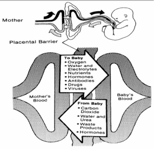
A few interesting points:
You will see that antibodies are small enough to cross the placenta. This gives the baby a passive immunity that can protect it for a short time from any pathogens it encounters.
Drugs such as alcohol and nicotine can cross the placenta. This is why it is so vital that pregnant mothers do not smoke and drink to ensure that the foetus’ development is not affected by these drugs.
