
Tagged: 3.8
Female Reproductive System: Grade 9 Understanding for IGCSE Biology 3.8
The human male and female reproductive systems are made from the same embryonic cells and are perhaps more similar in structure and function than is first apparent. There are two ovaries protected within the pelvic cavity. The ovary is the site of egg cell production. The egg cell is the female gamete and is haploid – it has only one chromosome from each homologous pair. The ovaries are also endocrine organs that produce the female sex hormones oestrogen and progesterone.
[Indeed differences between the gametes is the essential difference between male and female organisms. Females are always individuals who produce a small number of large, often immobile gametes. You can easily remember this: female – few, fixed, fat. Males are organisms that produce large numbers of small, motile games. Male – many, mini, motile.]
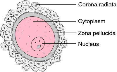
This diagram shows the human egg cell after it has been released from the ovary into the Fallopian tubes (or oviduct). The egg cell is coloured pink in the diagram above (if you are being picky it is not really an egg but a cell called a secondary oocyte but I won’t stress over this now…) The egg cell is surrounded by a thick jelly-like layer called the zona pellucida and then by a whole cluster of mother’s cells from her ovary – the corona radiata. The big idea to remember is that the egg cell is very large compared to sperm cells: it is one of the largest cells in humans with a diameter of about 500 micrometers.
The Fallopian tubes carry the egg down towards the uterus. The lining of the Fallopian tubes is covered in a ciliated epithelium. The cilia waft to generate a current that helps move the egg down towards the uterus. Sperm cells have to swim against this current to reach the egg in the tubes. The Fallopian tube is the usual site for fertilisation to occur.
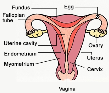
Once fertilisation has occurred, the newly formed zygote divides over and over again by mitosis to form a ball of cells called an embryo. The embryo continues its journey down the Fallopian tube until it reaches the uterus. The uterus (womb) is a muscular organ with a thickened and blood-rich lining called the endometrium. Implantation occurs when the embryo attaches to the endometrium and over time, a placenta forms. The embryo develops into a foetus and remains in the uterus for 9 months.
The cervix is a narrow opening between the uterus and the vagina. It holds the developing foetus in the uterus during pregnancy but dilates (widens) at birth to form part of the birth canal. The vagina is the organ into which sperm are deposited from the man’s penis during sexual intercourse. The lining of the vagina is acidic to protect against bacterial pathogens and the sperm cells released into the vagina quickly start to swim away from the acidity in grooves in the lining. These grooves lead to the cervix and hence into the uterus.

Male Reproductive Systems: Grade 9 Understanding for IGCSE Biology 3.8
I am slightly wary about writing about the male and female reproductive systems. Not because I get embarrassed with this topic (5 terms of human dissection at medical school removed any squeamishness about body parts….) But rather that I worry that the school’s internet filters might start blocking my website if the wrong words appear. But you don’t know until you try, so here goes…..
Male Reproductive System
I will start with the male reproductive system as males are simpler than females in many, many ways… The male reproductive system has three functions:
- to produce the male gametes, sperm cells, at a prodigious rate
- to make the male sex hormone testosterone
- to act as a delivery system to ensure sperm cells are carried into the female reproductive tract in conditions that will allow them to fertilise an egg
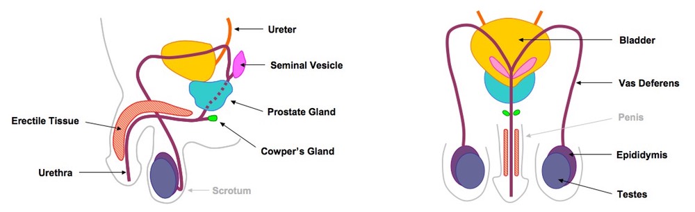
The first two functions listed above happen in the testis. There are cells in the testis that secrete the hormone testosterone into the blood from puberty onwards. Testosterone switches on secondary sexual characteristics in the male (body hair growth, muscle development, change in pitch of voice etc.) after puberty. The main part of the testis is made up of very long coiled tubules called seminiferous tubules in which the sperm cells are made.
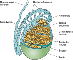
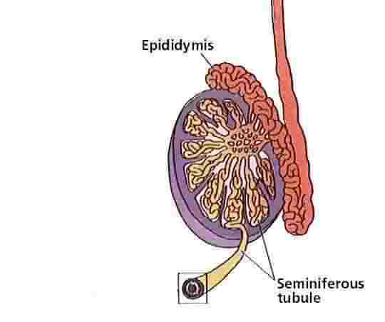
Humans have over a hundred meters of seminiferous tubules in total in both testes and this allows sperm cells to made at a very fast rate. Even though it takes around 75 days to make an individual sperm cell, the testes make them at a rate of around 85 million sperm cells per day. The epididymis is found next to the testis in the scrotum and is a coiled tube in which sperm cells continue to develop and mature. Sperm are stored here too in readiness for ejaculation.
Everyone knows that in humans the testes are found outside the body cavity in order to keep them cool. Sperm production happens at a maximal rate 3 degrees below core body temperature and having testes outside the body keeps them at this temperature.
The vas deferens is a tube lined with smooth muscle that carries sperm cells away from the testis for ejaculation. As you can see it loops around the back of the bladder, before joining up with the urethra just below the bladder. The urethra is the tube that carries urine away from the bladder but can also carry semen once the vas deferens has joined with it.
There are three accessory glands in the male system (prostate gland, seminal vesicle and the Cowpers‘ glands) These glands produce the fluid that when mixed with the sperm cells is called semen. Semen contains a sugar fructose to provide energy for the sperm cells to swim. It is slightly alkaline to neutralise the acidity in the vagina and also contains mucus to make the fluid easy to move along the tubes.
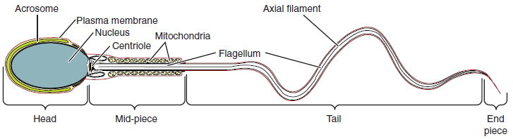
The sperm cells only acquire the ability to swim when in the epididymis and only become totally mature and able to fertilise the egg right next to the egg cell in the female tract.
The penis is an organ that contains erectile tissue that can fill with blood to allow the penis to fit into the female vagina for ejaculation.
