Tagged: cell

Cell Structure: Grade 9 Understanding for IGCSE Biology 2.2 2.3 2.4
All living organisms are made from cells. Indeed the cellular nature of life is one of the universal features shared by all life on earth. Some organisms are made from just one cell (unicellular organisms) while at some point around 1 billion years ago, cells starting clumping together and specialising to form multicellular organisms such as animals and plants.
What do all cells have in common?
All cells are surrounded by a cell membrane. The cell membrane is made from a mixture of proteins and a type of lipid called a phospholipid. The cell membrane serves many functions but perhaps the most significant is acting as a partially permeable barrier that can control which molecules can enter and leave the cell.
Inside the cell membrane there is a watery solution of chemicals called the cytoplasm. The cytoplasm is the site of many metabolic reactions in the cell because many enzymes are dissolved in the cytoplasm. The cytoplasm also contains many tiny nano machines for assembling proteins called ribosomes.
And that is about it for things all cells have in common. Prokaryote cells (bacteria) have a very different cell structure with no organelles but in this section you need to understand the simplified structure of two eukaryote cells: a typical animal (on the left below) and a typical plant cell (on the right).

Both animal and plant cells have a nucleus. This is the largest organelle and contains the DNA which is the genetic material. The DNA is found in long thread-like structures called chromosomes. The nucleus controls the division of the cell and also the various functions of the cell by regulating which proteins get made.

Animal and Plant cells both contain mitochondria which are the organelles associated with aerobic respiration. Mitochondria are recognisable in the cytoplasm of the cell as sausage-shaped organelles with a folded inner membrane (see diagram above).

Structures found only in Plant cells
1) All plant cells have a thick rigid cell wall made of the carbohydrate cellulose. The cell wall allows plant cells to become turgid since when the cell takes in water by osmosis, the rigid cell wall prevents the cell from bursting. The cell wall also acts as a transport pathway across plant tissues and can provide a barrier to some pathogens.
2) All plant cells have a large permanent central sap vacuole. This organelle is bounded by a membrane called the tonoplast and in many plant cells takes up the majority of the volume of the cell.

The sap vacuole provides a compartment in the cell into which excretory molecules can be moved to stop them poisoning the cytoplasm. It also plays a role in the water balance of plant cells since because of all the solute dissolved in it, the cell sap has a low water potential. This helps draw in water by osmosis from the cytoplasm and hence from outside the cell across the cell membrane.
3) Many but not all plant cells contain chloroplasts. These are organelles associated with the process of photosynthesis. Chloroplasts can be recognised in a light microscope image as small, green structures in the cell. The green pigment comes from the chlorophyll molecules that trap energy from sunlight. In an electron micrograph, chloroplasts are distinguished due to their stacks of membrane discs called grana.


Differences between plant and animal cells

Cell Differentiation and Specialised Cells: Grade 9 Understanding for IGCSE Biology 2.5B
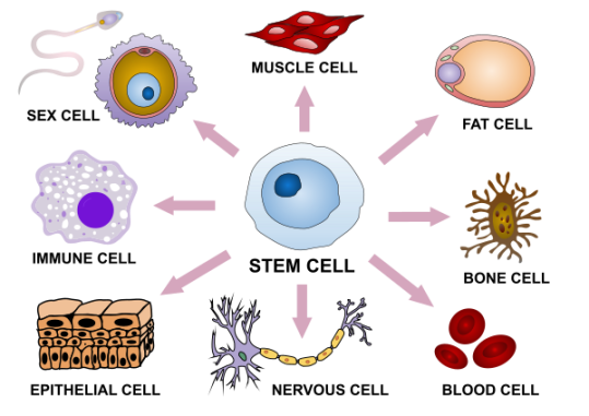
You will have learned at KS3 about the basic structure of a “typical” animal cell. But our bodies are not made of cells that look like this “typical” cell. Humans have just over 200 different types of cell, each specialised to carry out a particular function. For example red blood cells are specialised for transporting oxygen, muscle cells are specialised for movement and sperm cells are specialised as the male gamete for delivering a haploid nucleus to the egg cell.

The diagram above shows examples of a few types of specialised cells from the human body.
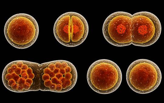
These specialised cells are produced in the process of cell division.
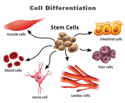
Cells that are not yet specialised but that retain the ability to develop into a variety of different cell types are called stem cells. Many cells in the embryo are stem cells (as they have not yet specialised into a particular cell type) but we also have a few stem cells in the adult (for example the cells in the bone marrow that can develop into all the different cell types in blood).
The process by which stem cells develop into specialised cells is called differentiation. Luckily you don’t need to understand exactly how this works but basic idea is this: differentiation involves certain genes in the nucleus being switched on and off so that a specialised cell only makes a certain set of proteins. Remember that a gene is a section of our DNA that codes for a single protein. Nerve cells make the proteins needed to send nerve impulses, white blood cells make the proteins needed to combat infections. You get the idea…..
Stem cells play an important role in medicine but that’s for another post……… If you want to read more about stem cells, this website is a good place to start.
Cell Structure: Grade 9 Understanding for iGCSE Biology 2.2 2.3 2.4
All living organisms are made from cells. Indeed the cellular nature of life is one of the universal features shared by all life on earth. Some organisms are made from just one cell (unicellular organisms) while at some point around 1 billion years ago, cells starting clumping together and specialising to form multicellular organisms such as animals and plants.
What do all cells have in common?
All cells are surrounded by a cell membrane. The cell membrane is made from a mixture of proteins and a type of lipid called a phospholipid. The cell membrane serves many functions but perhaps the most significant is acting as a partially permeable barrier that can control which molecules can enter and leave the cell.
Inside the cell membrane there is a watery solution of chemicals called the cytoplasm. The cytoplasm is the site of many metabolic reactions in the cell because many enzymes are dissolved in the cytoplasm. The cytoplasm also contains many tiny nano machines for assembling proteins called ribosomes.
And that is about it for things all cells have in common. Prokaryote cells (bacteria) have a very different cell structure with no organelles but in this section you need to understand the simplified structure of two eukaryote cells: a typical animal (on the left below) and a typical plant cell (on the right).
Both animal and plant cells have a nucleus. This is the largest organelle and contains the DNA which is the genetic material. The DNA is found in long thread-like structures called chromosomes. The nucleus controls the division of the cell and also the various functions of the cell by regulating which proteins get made.
Animal and Plant cells both contain mitochondria which are the organelles associated with aerobic respiration. Mitochondria are recognisable in the cytoplasm of the cell as sausage-shaped organelles with a folded inner membrane (see diagram above).
Structures found only in Plant cells
1) All plant cells have a thick rigid cell wall made of the carbohydrate cellulose. The cell wall allows plant cells to become turgid since when the cell takes in water by osmosis, the rigid cell wall prevents the cell from bursting. The cell wall also acts as a transport pathway across plant tissues and can provide a barrier to some pathogens.
2) All plant cells have a large permanent central sap vacuole. This organelle is bounded by a membrane called the tonoplast and in many plant cells takes up the majority of the volume of the cell.
The sap vacuole provides a compartment in the cell into which excretory molecules can be moved to stop them poisoning the cytoplasm. It also plays a role in the water balance of plant cells since because of all the solute dissolved in it, the cell sap has a low water potential. This helps draw in water by osmosis from the cytoplasm and hence from outside the cell across the cell membrane.
3) Many but not all plant cells contain chloroplasts. These are organelles associated with the process of photosynthesis. Chloroplasts can be recognised in a light microscope image as small, green structures in the cell. The green pigment comes from the chlorophyll molecules that trap energy from sunlight. In an electron micrograph, chloroplasts are distinguished due to their stacks of membrane discs called grana.
Differences between plant and animal cells

Levels of Organisation: Grade 9 Understanding for IGCSE Biology 2.1
The Easter holiday is the most important time of year for this iGCSE Biology blog. With exams in early May, the next few weeks should be the time when students are working at their maximal rate. I intend to add one post a day such that by the middle of April, the entire EdExcel iGCSE Biology specification has been covered on this blog. This should then make it a useful resource for all GCSE Biology students to help them with their revision.
Today I will put up two posts that will look at two of the simplest topics in the specification: the first one will be Levels of Organisation and then Cell Structure (2.1, 2.2, 2.3 and 2.4)
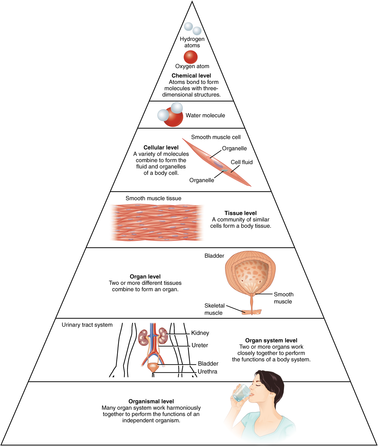
Living things (or organisms to be precise) are complex entities. Even the simplest organism will be made up of millions of different molecules arranged in an organised and complex way. Human beings are organisms made up of about 10 trillion cells of roughly 210 different cell types all put together in a organised and systematic way. It makes it much easier to study such complexity if we have a system to break the complexity down into constituent parts. This is what scientists mean by levels of organisation.
So, starting with the smallest things that might be of interest to a biologist……
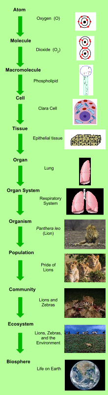 All matter on earth including the matter of living things is made of atoms (e.g. a carbon atom, an oxygen atom etc.). Atoms can combine together in a variety of ways to form molecules (a water molecule H2O, a carbon dioxide molecule CO2,) How atoms combine to form molecules is chemistry, and the levels of organisation smaller than an atom forms part of physics, so we won’t worry too much about them….
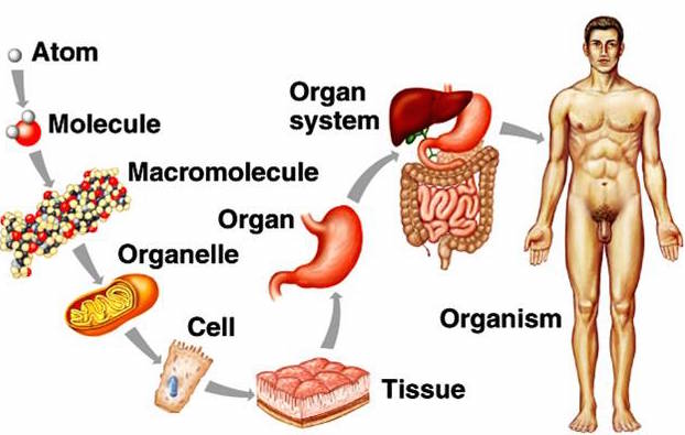
But molecules in an organism are interesting and worth studying – you learn about carbohydrates, lipids, proteins, DNA in your iGCSE course. These molecules can be grouped together to form structures inside cells called organelles. If you are being really precise with your terminology, an organelle is a membrane-bound compartment inside a eukaryotic cell (remember bacterial cells have no organelles at all). Examples of organelles are structures like the nucleus, chloroplasts, mitochondria and so on.
Cells are structures enclosed by a cell membrane that contain many different organelles. You have probably looked at a human cheek cell using a light microscope at some point in the past. In multicellular organisms, cells of the same type are often attached together to form a Tissue. A tissue is a group of similar cells often attached to each other that carry out the same function in an organism. (It is also a small disposable piece of rectangular fabric used for blowing your nose but that is something quite different….) Tissues are grouped together to form larger structures called Organs. For example, the lungs are an organ made up of a particular arrangement of epithelial tissues together with some blood and connective tissues. Organs can be grouped into Organ Systems based on their function such as the Digestive System (oesophagus, tongue, stomach, pancreas, liver, intestines etc.) An Organism such as you or I is made up of many organ systems (nervous system, cardiovascular system, digestive system, excretory system and so on….. You get the idea I’m sure!)
You can study levels of organisation bigger than the organism. This branch of biology is called Ecology – and indeed you should know the meaning of the terms population, community and ecosystem – but perhaps that is for another post……

Blood part 2 White Blood Cells – Grade 9 Understanding for GCSE Biology 2.59 2.62 2.63B
The previous post looked at the structure and function of red blood cells and plasma. Now it is time to turn our attention to the rather more complex topic of white blood cells….. This is a topic in which the complexity can put people off but I am deliberately going to keep things simple (I hope!). If you are thinking about revision for GCSE, don’t worry about anything more complicated than in this post.

There are many types of white blood cell found in blood. But let’s keep things simple…. You need to understand the role of lymphocytes and phagocytes in defending the body against pathogens.
A pathogen is defined as “a microorganism that can cause a disease” and pathogens may be bacteria, viruses, protistans or fungi. Can you give me an example of an infectious disease caused by each class of pathogen?
The structure of these two classes of white blood cell is important. The commonest phagocytes in blood are called neutrophils and they are easily recognised by their irregular shaped nucleus and cytoplasm packed full of granules. Lymphocytes are much smaller white cells and are identifiable by their clear cytoplasm and large spherical nucleus that takes up 90% of the volume of the cell.


So now we should look at how these two types of white blood cells defend the body against pathogens. Remember that the account on this post is an over-simplification of what is in reality an extremely complex process.
Let’s start with a phagocyte. These large cells are able to engulf invading pathogens in the blood and tissue fluid by a process called phagocytosis.

The phagocyte pushes out projections of its cytoplasm around the clump of bacteria. These projections are called pseudopodia and when they meet, the cell membrane of the phagocyte fuses together leaving the bacteria enclosed in a tiny membrane packet called a vesicle inside the cytoplasm. The phagocyte then fuses other vesicles that contain powerful digestive enzymes with the vesicle with the bacteria in, leading to the death and destruction of the bacteria. Simple.
The problem for phagocytes is this: how do they know what to engulf and destroy? This is where lymphocytes come in. One class of lymphocyte is able to secrete small soluble proteins called antibodies into the blood. Antibodies are specific to a particular surface marker on the invading pathogen and bind to it because the shape of the antibody and the shape of the surface marker are complimentary.

Now people always get confused between antibodies (the small soluble Y-shaped proteins secreted by lymphocytes) and antigens (the surface markers on the invading pathogen). Make sure you are completely clear on the difference in meaning of these two words….
 This diagram shows antibodies (green) binding to surface markers (antigens) on a bacterial cell.
Antibodies produced by lymphocytes will coat the invading pathogen by binding to antigens on its surface. One effect of this is that phagocytes are stimulated to engulf the antibody-coated organism.
There are many different types of lymphocyte and not all can produce antibodies. Another important function of lymphocytes is to kill your own body cells when they are corrupted, either by the presence of a virus or by becoming cancerous.
Finally, can I draw your attention to two previous posts linked to this one. The first is on the role of platelets in blood clotting, the second on the difficult topic of immunity and how lymphocytes are responsible for giving you lifelong protection against certain infectious diseases.
https://pmgbiology.wordpress.com/2014/04/07/immunity-a-understanding-for-biology-igcse/
As always, please ask me questions either via the comment section below the post or with a tweet…. I will do my best to respond to any questions from anyone who is bothered to read my posts!
Blood part 1 Plasma and RBCs: Grade 9 Understanding for IGCSE Biology 2.59, 2.60, 2.61
Blood is a tissue in the body that plays a variety of roles in transport and in defending the body against disease. It is an unusual tissue since it is a liquid, with many different kinds of cells suspended in a watery solution called plasma. 
Plasma makes up 55% of the volume of blood and is a solution of many different chemicals in water. For example, the plasma contains dissolved glucose, amino acids and other products of digestion from the intestines. It also transports the waste molecule urea from the liver where it is made to the kidney where it is excreted. Blood plasma contains dissolved carbon dioxide, mostly in the form of hydrogencarbonate ions. Many hormones (for example testosterone, ADH, adrenalin) are transported in the blood plasma and because the plasma is mostly water, it provides a good way of moving heat around the body from respiring muscles to the skin where it can be lost.
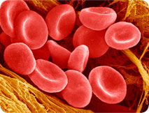
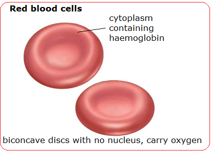
The most common cell in blood are the red blood cells (or erythrocytes). These tiny cells are adapted for the transport of oxygen. Each red blood cell contains around 270 million molecules of a transport protein, haemoglobin. Each molecule of haemoglobin can bind up to four molecules of oxygen in the lungs and then unload the oxygen when the red blood cell passes through a capillary in an actively respiring tissue.

(Don’t worry too much about the structure of the protein – this is A level stuff really…. Just remember haemoglobin is a transport protein for oxygen found in red blood cells)
As well as being packed full of haemoglobin molecules, red blood cells have other adaptations for transporting oxygen. Red blood cells lose their nucleus during their development as this allows more haemoglobin to be packed into each cell. Having no nucleus means the red blood cell cannot divide nor repair damage to its structure. This is why each red blood cell only lives for 100-120 days in the body.
Red blood cells have a characteristic shape. It is called a biconcave disc and they have an especially flexible shape. Remember that a capillary is actually smaller in diameter than a red blood cell, so the cells have to squeeze through capillaries in single file…..