
Tagged: heart
“How to build a human heart” video
Adrenaline: Grade 9 Understanding for IGCSE Biology 2.94
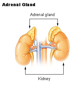
Adrenaline is a hormone produced in the adrenal glands which are found on top of the kidneys in the abdomen.
A hormone is “a chemical released by a specialised gland called an endocrine gland into the bloodstream. The hormone travels around the body in the blood plasma and then causes an effect elsewhere in the body by binding to receptors found on certain target cells”.
You should know some other examples of hormones – testosterone, oestrogen, progesterone, ADH – to name a few. Please learn this definition too: it would be wonderful if you got a 3 mark question asking you to define a hormone….

There are many cells in the body that contain receptors for adrenaline. This allows the hormone to exert an effect on a wide variety of tissues. For example there are adrenaline receptors in the pacemaker of the heart and adrenaline will cause the heart to beat faster (more beats per minute) and also with more force.
When is adrenaline released by the adrenal glands into the blood?
Adrenaline is secreted into the blood in times of danger or stress. It prepares the body to either run away from the danger or indeed to battle against it. For this reason, adrenaline is often described as a “fight or flight” hormone.
What are some of the effects of adrenaline?
Target Tissue Effect
Heart Increase in heart rate, increase in cardiac output
Lungs Bronchioles dilate (widen)
Muscles Arteries in muscle dilate to allow more blood to flow to muscles
Skin/Digestive system Arteries in skin/digestive system constrict so less blood flows
Liver Liver breaks down glycogen into glucose to raise blood glucose conc.
Iris Radial muscles in iris contract causing pupil dilation
The overall effect is that the skeletal muscles are supplied with more oxygen and more glucose so they can respire aerobically. This allows the muscle to contract more efficiently.

Cardiac cycle and the Human Heart: Grade 9 Understanding for IGCSE Biology 2.65 2.66
The human heart is an organ found in the middle of the thorax. It is made from a specialised type of muscle called cardiac muscle and acts as a pump to push blood around the two circulatory systems. In fact it is better to think of the heart as two separate pumps, one for the pulmonary circulatory system to the lungs and one for the systemic system that supplies blood to all the other body organs.
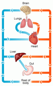
The diagram above shows in a simplified way this double circulatory system. The lungs are supplied with deoxygenated blood direct from the heart in the pulmonary artery but when the blood has passed through the capillaries in the lungs, it travels back to the heart in the pulmonary vein before being pumped in the systemic system around the body. Although cardiac muscle looks similar to the muscle that attaches to bones and moves the skeleton, it differs functionally in one important way. Cardiac muscle is myogenic: this means that the muscle fibres will contract without the need for a nerve impulse from the brain to initiate the contraction. Incidentally this is why a heart transplant is a possible surgical procedure. A transplanted heart will beat happily in the new body even though all the nerves going to the heart will have been cut in the surgery. You cannot have a biceps transplant at the moment because the transplanted biceps muscle would not do anything in the new patient. For the transplanted biceps to contract, the millions of individual neurones going to it would need linking up individually and this is not possible.

This is a simplified diagram showing the structure of the heart. You can see that the left and right sides of the heart are completely separate from each other. This is essential because the right side of the heart contains deoxygenated blood and the left side oxygenated blood. There are four chambers in the heart: two small atria at the top, and two larger ventricles at the bottom. The atria collect blood from the veins, the ventricles pump blood out of the heart into arteries.
There are four sets of valves in the heart: the easiest way to remember where they are is to think that blood has to pass through a set of valves as it leaves each chamber. Valves allow blood to flow through them in one direction only. The AV valves stop blood going back from the ventricle into the atrium when the ventricle contracts, the aortic and pulmonary valves (not labelled on the diagram above for some reason….) prevent blood falling back into the ventricles in between heart beats.
You also need to know the four main blood vessels that are attached to the heart. The vena cava is the largest vein in the body and carries deoxygenated blood from the organs of the body into the right atrium. The pulmonary artery comes out of the right ventricle and pumps this deoxygenated blood to the lungs. Oxygenated blood returns from the lungs to the left atrium in the pulmonary veins, passes into the left ventricle and is then pumped out of the heart in the aorta, the biggest artery in the body.

NB I strongly suggest you do not use the terms bicuspid, mitral or tricuspid valve to label the valves between the atria and ventricles… In an exam it is easy to get these similar words muddled, so I would always call the valve between the left atrium and left ventricle the left atrio-ventricular valve (never bicuspid or mitral valve) and I would always call the valve between the right atrium and right ventricle the right atrio-ventricular valve (never tricuspid). I know many of you will ignore this advice but it’s good to get it off my chest……
I’ve just spent 45 minutes trying to find a good video on heart structure to put into the post but without success…. If anyone knows a really good YouTube clip (must be under 5 minutes) please add a link as a comment at the foot of this post.
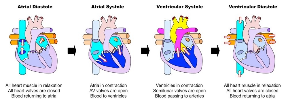
Events of the Cardiac Cycle
This is a topic that requires some simplification as it is easy to get confused. The Cardiac Cycle is simply a word for the sequence of events that happen in a heart beat. At the simplest level, the cardiac cycle consists of three phases:
1. Diastole. During this stage the cardiac muscle is relaxed (the heart is between beats) and blood can enter the atria and then fall into the ventricles through the open AV valves.
2. Atrial Systole. This stage in when the cardiac muscle in the atria contract, increasing the atrial pressure and pushing blood down into the ventricles. There is a small region in the wall of the right atrium called the sino-atrial node (or pacemaker) which initiates each heart beat.
3. Ventricular Systole. After a short delay, the cardiac muscle in the ventricles contracts. This increases the blood pressure in the ventricle which in turn causes the AV valve to close, and the aortic or pulmonary semilunar valves to open. As these valves at the exit of the ventricle open, blood gets pushed into the arteries and out of the heart.
Look at the diagram above and make sure you understand the meaning of these three terms.
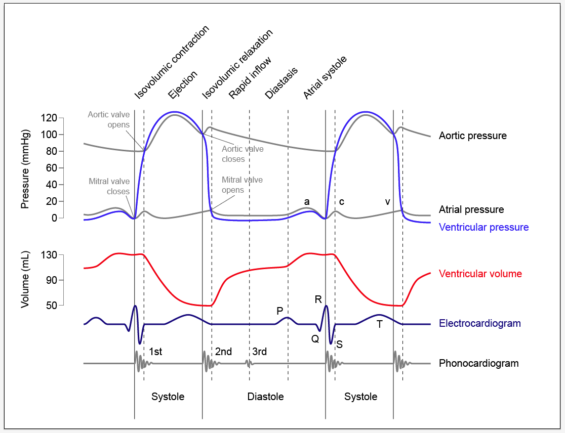
The diagram below is much more complex and perhaps you should not worry too much about it….. The important bit is to understand when the valves in the heart open and when they close. It is quite simple really although you would be amazed how confused people can get….
The opening and closing of a valve is not “controlled” in any meaningful way in the heart. The valve has a structure that will only allow it to open in one direction. Let’s consider the AV valves. These can open to allow blood to pass from the atria into the ventricles during atrial systole but will close during ventricular systole to stop the blood flowing back where it came from.
The AV valve will be open whenever the blood pressure in the atria is greater than in the ventricle.
The AV valve will be closed whenever the blood pressure in the ventricle is greater than in the atrium.
Look at the graph of pressure changes below. You can see the mitral valve (I hate that name) is closed as soon as the ventricular pressure exceeds the atrial pressure and it opens again during ventricular diastole as the pressure in the ventricle drops.

Diseases associated with Smoking: Grade 9 Understanding for IGCSE Biology 2.49 2.67
The health risks of smoking cigarettes and other tobacco products are well understood. But in iGCSE exams, it can sometimes be difficult to work out exactly how much detail the examiner wants, especially on an open-ended question. This blog post is an attempt to demonstrate some of the key areas of understanding that you should be aiming to show in your answers.

Cigarettes contain a wide variety of toxic chemicals: you should focus your understanding on three examples.
Nicotine is the stimulant drug found in cigarette smoke and also the reason smokers can easily become dependent on smoking. Nicotine is rapidly absorbed into the blood in the lungs and it causes an increase in heart rate, an increase in blood pressure and the release of the hormone adrenaline (which has similar effects).
Carbon Monoxide is a poisonous gas that is produced whenever there is incomplete combustion of biological material. Carbon monoxide binds to haemoglobin in the red blood cells in place of oxygen and so the smoker will be transporting less oxygen in her blood, and this leads to a whole load of implications for health.
Tar is the name given to a large number of different chemicals found in cigarette smoke. Tar forms droplets in the smoke which can condense in the airways and alveoli. Many of the chemicals in Tar are carcinogens – that means they promote the formation of cancer. Tar also damages the cilia in the trachea and bronchi and makes the thin walls of the alveoli lose their elasticity and so damage more easily.
Diseases of the Lungs associated with Smoking

1) Emphysema
Emphysema is a disease in which the thin walls of the alveoli break down. Tar droplets condense onto the alveolar wall, making them rigid and inflexible. Smokers are coughing a great deal to remove the mucus from their lungs and this coughing can cause the sticky, rigid tar coated alveolar wall to degenerate.
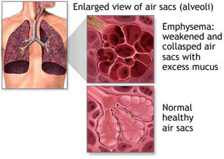

A patient with emphysema will have a greatly reduced surface area for gas exchange due to all the collapsed alveoli. So the diffusion of oxygen into the blood would be reduced causing breathlessness and impacting health.
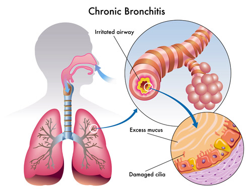
2) Chronic Bronchitis
The bronchi can become inflamed and narrowed due to the overproduction of mucus and the fact that the muco-ciliary escalator does not function to waft mucus up the bronchi. A narrowed bronchus makes breathing harder and compounds the problems of emphysema described above. It also promotes coughing which damages the alveoli still further.
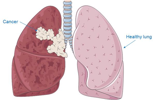
3) Lung cancer
Carcinogens in cigarette smoke can make it much more likely that cells in the lungs and airways develop mutations that lead to cancer. Cancer is a disease in which cells start to divide out of control to form a tumour and then cells in the tumour break off and travel round the bloodstream to form secondary tumours elsewhere in the body. Lung cancers can be hard to treat and are a common cause of early death in cigarette smokers.
Diseases of the Cardiovascular System associated with Smoking
It is not just the lungs that are affected in patients who smoke. Cigarette smoking is a significant risk factor in the commonest cause of death in the UK: coronary heart disease (CHD)
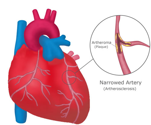
CHD is a disease in which the arteries that supply the cardiac muscle in the heart (the coronary arteries) get narrowed due to the build up of fatty plaque in their walls. This is a condition called atherosclerosis. A narrowed coronary artery means that an area of cardiac muscle is starved of oxygen and so can die. This may interfere with the electrical coordination of the heart beat causing a cardiac arrest or heart attack.
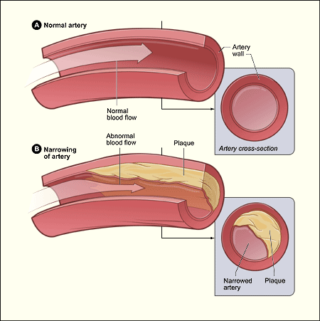
Patients who smoke have a higher blood pressure than normal due to the stimulant effects of nicotine. A high blood pressure makes the early damage to the arterial lining more likely as well as making the blood more likely to clot. Blood clots form in the final stages of the disease around the plaque and can complete block the blood vessel. CHD is a major cause of death across the world and it is well known that stopping smoking can do a great deal to lower a patient’s risk of this potentially deadly disease.
Human Transport IGCSE – a few pointers for Grade 9 Understanding 2.59, 2.63B, 2.69
I have had a request from a student to write about the level of details needed in the section of the specification on human transport. Here are the relevant bullet points from the specification, together with a very brief outline of the kinds of details to learn:
- Blood composition 55% plasma, 45% cells (red blood cells, white blood cells and platelets)
- Plasma functions – transport of dissolved carbon dioxide, dissolved glucose, urea, salts etc.and transport of heat around body
- Red Blood cells – no nucleus, each cell packed full of 250,000 molecules of haemoglobin, biconcave disc shape to squeeze through narrow capillaries
- Phagocytes/Lymphocytes – two types of white blood cell, phagocytes engulf foreign organisms in blood by phagocytosis, lymphocytes do many functions in defending the body against disease but many produce antibodies
- Vaccination with reference to memory cells and primary v secondary response (see below)
- Functions of clotting and role of platelets (prevent infection, stop blood loss – platelets play central role in clotting as they produce chemicals that are needed for clotting cascade
- Structure and function of the heart (learn names of chambers, blood vessels, names of four sets of valves and what they do)
- Role of adrenaline in changing heart rate during exercise (speeds it up to maximise cardiac output to muscles)
- Structure and functions of arteries/veins/capillaries (simple bookwork)
- General plan of circulation including heart, lungs, liver and kidneys (see below)
The two sections that are perhaps hardest to interpret are the ones on vaccination and the general plan of the circulation.
1) Key terms in vaccination to understand:
- Antigen
- Antibody
- Lymphocyte
- Clonal Selection theory
- Memory cells
- Effector cells (plasma cells)
- Primary response
- Secondary response
At the end of the process, you should be able to provide a clear concise answer to the following question?
Why is it that the first time your body encounters measles virus, you suffer from the disease measles? Why will someone who has had measles as a baby (or been immunised against it) never contract the disease measles even though the virus might get into their body many subsequent times?
2) The blood vessels involved in the four organs mentioned are described below.
Heart – receives blood from the coronary arteries which branch off the aorta before it has even left the heart: Why doesn’t the cardiac muscle in the heart just get the oxygen and nutrients it needs from the blood in the chambers?
Lungs – pulmonary artery takes blood from right ventricle to the lungs, pulmonary vein return oxygenated blood to the heart and empty it into the left atrium. What is unique about the composition of the blood in the pulmonary artery?
Liver – has a most unusual blood supply. There is a hepatic artery that branches off the aorta and brings oxygenated blood to the liver. Blood also goes to the liver in the hepatic portal vein which brings blood from the small intestine. Blood in the hepatic portal vein will contain lots of dissolved glucose and amino acids, both of which are processed in the liver. Deoxygenated blood leaves the liver in the hepatic vein. Find a diagram to show the arrangement of these three blood vessels.
Kidney – straightforward blood supply in that there is a renal artery and a renal vein. (important idea is that the renal artery is much much bigger than you would expect from the size of the organs: 25% of the cardiac output of blood flows through the kidneys on each circuit) Why do you think this is?
I hope this helps – more to follow when I get home from my holidays tomorrow afternoon…..
