Cardiac cycle and the Human Heart: Grade 9 Understanding for IGCSE Biology 2.65 2.66
The human heart is an organ found in the middle of the thorax. It is made from a specialised type of muscle called cardiac muscle and acts as a pump to push blood around the two circulatory systems. In fact it is better to think of the heart as two separate pumps, one for the pulmonary circulatory system to the lungs and one for the systemic system that supplies blood to all the other body organs.
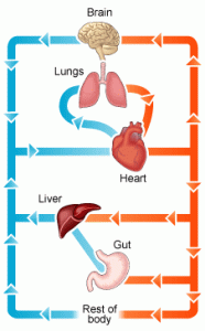
The diagram above shows in a simplified way this double circulatory system. The lungs are supplied with deoxygenated blood direct from the heart in the pulmonary artery but when the blood has passed through the capillaries in the lungs, it travels back to the heart in the pulmonary vein before being pumped in the systemic system around the body. Although cardiac muscle looks similar to the muscle that attaches to bones and moves the skeleton, it differs functionally in one important way. Cardiac muscle is myogenic: this means that the muscle fibres will contract without the need for a nerve impulse from the brain to initiate the contraction. Incidentally this is why a heart transplant is a possible surgical procedure. A transplanted heart will beat happily in the new body even though all the nerves going to the heart will have been cut in the surgery. You cannot have a biceps transplant at the moment because the transplanted biceps muscle would not do anything in the new patient. For the transplanted biceps to contract, the millions of individual neurones going to it would need linking up individually and this is not possible.

This is a simplified diagram showing the structure of the heart. You can see that the left and right sides of the heart are completely separate from each other. This is essential because the right side of the heart contains deoxygenated blood and the left side oxygenated blood. There are four chambers in the heart: two small atria at the top, and two larger ventricles at the bottom. The atria collect blood from the veins, the ventricles pump blood out of the heart into arteries.
There are four sets of valves in the heart: the easiest way to remember where they are is to think that blood has to pass through a set of valves as it leaves each chamber. Valves allow blood to flow through them in one direction only. The AV valves stop blood going back from the ventricle into the atrium when the ventricle contracts, the aortic and pulmonary valves (not labelled on the diagram above for some reason….) prevent blood falling back into the ventricles in between heart beats.
You also need to know the four main blood vessels that are attached to the heart. The vena cava is the largest vein in the body and carries deoxygenated blood from the organs of the body into the right atrium. The pulmonary artery comes out of the right ventricle and pumps this deoxygenated blood to the lungs. Oxygenated blood returns from the lungs to the left atrium in the pulmonary veins, passes into the left ventricle and is then pumped out of the heart in the aorta, the biggest artery in the body.

NB I strongly suggest you do not use the terms bicuspid, mitral or tricuspid valve to label the valves between the atria and ventricles… In an exam it is easy to get these similar words muddled, so I would always call the valve between the left atrium and left ventricle the left atrio-ventricular valve (never bicuspid or mitral valve) and I would always call the valve between the right atrium and right ventricle the right atrio-ventricular valve (never tricuspid). I know many of you will ignore this advice but it’s good to get it off my chest……
I’ve just spent 45 minutes trying to find a good video on heart structure to put into the post but without success…. If anyone knows a really good YouTube clip (must be under 5 minutes) please add a link as a comment at the foot of this post.
Events of the Cardiac Cycle
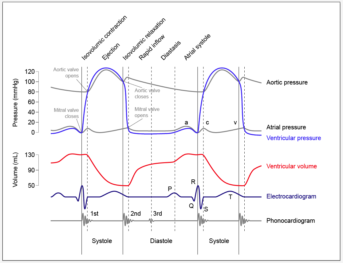
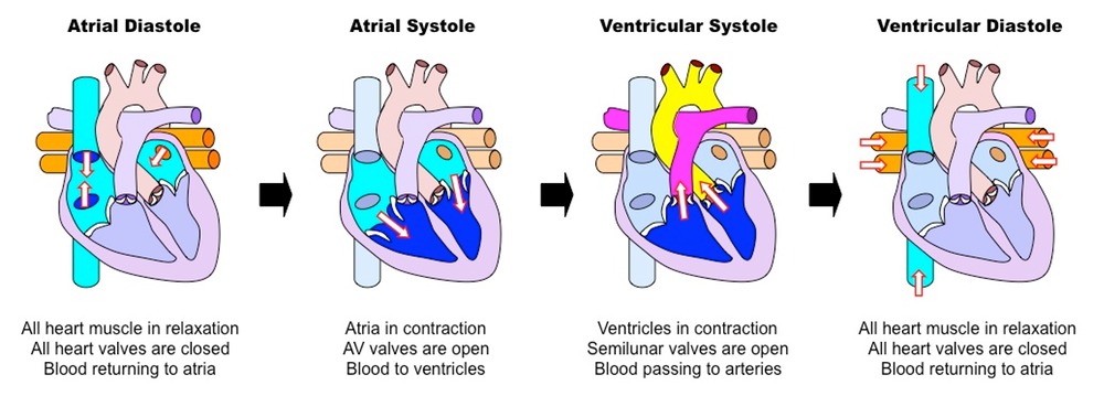
This is a topic that requires some simplification as it is easy to get confused. The Cardiac Cycle is simply a word for the sequence of events that happen in a heart beat. At the simplest level, the cardiac cycle consists of three phases:
1. Diastole. During this stage the cardiac muscle is relaxed (the heart is between beats) and blood can enter the atria and then fall into the ventricles through the open AV valves.
2. Atrial Systole. This stage in when the cardiac muscle in the atria contract, increasing the atrial pressure and pushing blood down into the ventricles. There is a small region in the wall of the right atrium called the sino-atrial node (or pacemaker) which initiates each heart beat.
3. Ventricular Systole. After a short delay, the cardiac muscle in the ventricles contracts. This increases the blood pressure in the ventricle which in turn causes the AV valve to close, and the aortic or pulmonary semilunar valves to open. As these valves at the exit of the ventricle open, blood gets pushed into the arteries and out of the heart.
Look at the diagram above and make sure you understand the meaning of these three terms.
The diagram below is much more complex and perhaps you should not worry too much about it….. The important bit is to understand when the valves in the heart open and when they close. It is quite simple really although you would be amazed how confused people can get….
The opening and closing of a valve is not “controlled” in any meaningful way in the heart. The valve has a structure that will only allow it to open in one direction. Let’s consider the AV valves. These can open to allow blood to pass from the atria into the ventricles during atrial systole but will close during ventricular systole to stop the blood flowing back where it came from.
The AV valve will be open whenever the blood pressure in the atria is greater than in the ventricle.
The AV valve will be closed whenever the blood pressure in the ventricle is greater than in the atrium.
Look at the graph of pressure changes below. You can see the mitral valve (I hate that name) is closed as soon as the ventricular pressure exceeds the atrial pressure and it opens again during ventricular diastole as the pressure in the ventricle drops.