
Category: Section 3: Reproduction and Inheritance
Cell Division part 2: Grade 9 Understanding of Mitosis for IGCSE 3.28 3.29
I thought I would make a video to explain the details of mitosis rather than typing up a blog post.
I would welcome any feedback on either of these videos. Do you find them useful? Do you prefer written blog posts? Leave a comment below if you want to let me know…..
Cell division video: a revision video for DNA structure and chromosomes
This is a summary video that might help those of you still struggling to get to grips with chromosomes and genes. I apologise for the terribly amateur production values on the video but hope the biological content at least might be useful….
Cell Division part I: Grade 9 Understanding for IGCSE 3.15, 3.28, 3.29
At the beginning of March each year, I get my Y11 classes to draw up a list of topics they want to go through again in revision. Cell Division is always there and it is not difficult to see why. Mitosis doesn’t make any sense unless you understand the concept of homologous pairs of chromosomes and I think you already know that very few iGCSE students do…… (Please see the various posts and videos on the blog on this topic before attempting to understand mitosis)
But there is actually very little to fear in the topic of cell division. If your teacher has told you about the various stages of mitosis that’s fine but you will not be asked to recall them in the exam, at least not if you are studying EdExcel iGCSE. So in this post I am going to try to focus on the key bits of understanding you need rather than bombarding you with unnecessary details.
1 Chromosomes come in pairs
This is the main idea you need before you start. In almost all sexually-reproducing organisms the cells are DIPLOID. This means that however many different sized chromosomes they have, in each cell there will be pairs of chromosomes (called homologous pairs)
So human cells contain 23 pairs of chromosomes (46 in total)
Remember that the number 46 only applies to humans. Other species have very different numbers of chromosomes in each cell (see table below)
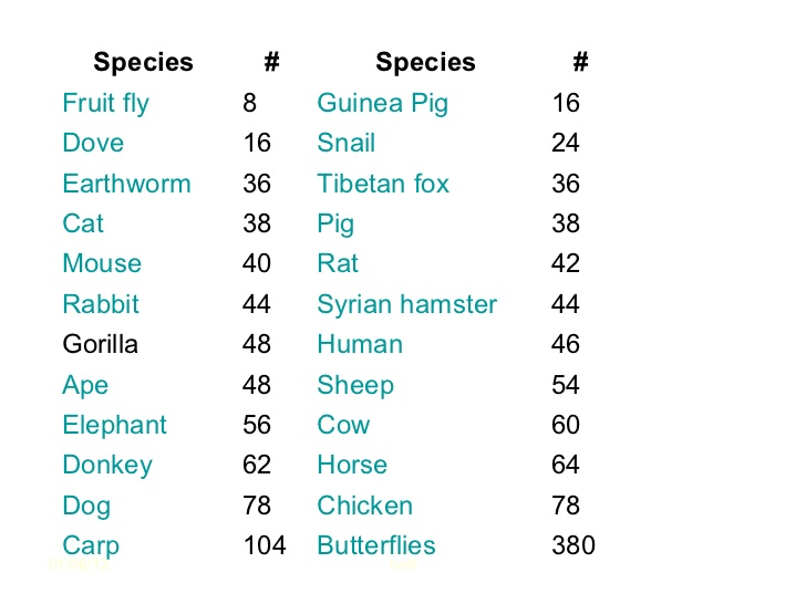
So doves have 8 pairs of chromosomes, dogs have 39 pairs of chromosomes, rats 21 pairs of chromosomes. The important point is not how many pairs each organism has but that they all have chromosomes that come in pairs!
The chromosomes any individual possesses is determined at the moment of fertilisation. Sperm and Egg cells (gametes) do not have pairs of chromosomes. They are the only cells in the body that are not diploid. Gametes only have one member of each pair of chromosomes. Cells which only have one member of each pair of chromosomes are called HAPLOID cells.
So every cell in the body is diploid and genetically identical apart from the gametes which are haploid.
2 Organisms that reproduce sexually need two different types of cell division

The fertilised egg (zygote) is a diploid cell. It has pairs of chromosomes that originate one from each parent via the gametes. Every cell division in growth and development of the embryo and foetus until birth, every cell division in growth and repair after birth always produces two genetically identical and diploid cells from the one original cell. This cell division that produces genetically identical diploid cells is called Mitosis.
Gametes (sperm and egg cells) need to be made by a different process. If gametes were diploid then there would be a doubling of the chromosome number every generation and that clearly wouldn’t do. So a different way of dividing the nucleus has evolved. It doesn’t produce genetically identical diploid cells but produces gametes that are haploid and genetically unique. This process is called Meiosis and is only used in the production of gametes.
3 Mitosis is involved in growth, repair, asexual reproduction and cloning
Any process in the body in which the outcome required is the production of genetically identical diploid cells will use mitosis. (It is not too complicated an idea to see that if you don’t need to make gametes and fuse them together in fertilisation, you can just copy cells by mitosis over and over again. All the daughter cells will be exact copies of each other and diploid.
Now I know this post is not going to satisfy everyone. I know some of you will want to read about the cell cycle, prophase, metaphase, centrioles, spindle fibres and the condensation of chromosomes, chromatids being pulled apart etc. etc.) And just for you, I will write a post later today on the details of Mitosis….. But please remember that if you are using the blog to revise for exams, none of this second post is necessary and none of it will be tested in the Edexcel iGCSE paper. If you are doing revision, focus on the key understanding ideas discussed above. And as always, please leave a reply below to ask questions, comment or leave feedback – all comments welcome!
Female Reproductive System: Grade 9 Understanding for IGCSE Biology 3.8
The human male and female reproductive systems are made from the same embryonic cells and are perhaps more similar in structure and function than is first apparent. There are two ovaries protected within the pelvic cavity. The ovary is the site of egg cell production. The egg cell is the female gamete and is haploid – it has only one chromosome from each homologous pair. The ovaries are also endocrine organs that produce the female sex hormones oestrogen and progesterone.
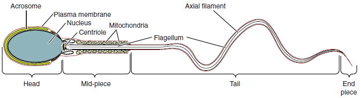
[Indeed differences between the gametes is the essential difference between male and female organisms. Females are always individuals who produce a small number of large, often immobile gametes. You can easily remember this: female – few, fixed, fat. Males are organisms that produce large numbers of small, motile games. Male – many, mini, motile.]
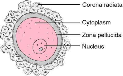
This diagram shows the human egg cell after it has been released from the ovary into the Fallopian tubes (or oviduct). The egg cell is coloured pink in the diagram above (if you are being picky it is not really an egg but a cell called a secondary oocyte but I won’t stress over this now…) The egg cell is surrounded by a thick jelly-like layer called the zona pellucida and then by a whole cluster of mother’s cells from her ovary – the corona radiata. The big idea to remember is that the egg cell is very large compared to sperm cells: it is one of the largest cells in humans with a diameter of about 500 micrometers.
The Fallopian tubes carry the egg down towards the uterus. The lining of the Fallopian tubes is covered in a ciliated epithelium. The cilia waft to generate a current that helps move the egg down towards the uterus. Sperm cells have to swim against this current to reach the egg in the tubes. The Fallopian tube is the usual site for fertilisation to occur.
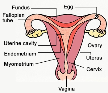
Once fertilisation has occurred, the newly formed zygote divides over and over again by mitosis to form a ball of cells called an embryo. The embryo continues its journey down the Fallopian tube until it reaches the uterus. The uterus (womb) is a muscular organ with a thickened and blood-rich lining called the endometrium. Implantation occurs when the embryo attaches to the endometrium and over time, a placenta forms. The embryo develops into a foetus and remains in the uterus for 9 months.
The cervix is a narrow opening between the uterus and the vagina. It holds the developing foetus in the uterus during pregnancy but dilates (widens) at birth to form part of the birth canal. The vagina is the organ into which sperm are deposited from the man’s penis during sexual intercourse. The lining of the vagina is acidic to protect against bacterial pathogens and the sperm cells released into the vagina quickly start to swim away from the acidity in grooves in the lining. These grooves lead to the cervix and hence into the uterus.

Male Reproductive Systems: Grade 9 Understanding for IGCSE Biology 3.8
I am slightly wary about writing about the male and female reproductive systems. Not because I get embarrassed with this topic (5 terms of human dissection at medical school removed any squeamishness about body parts….) But rather that I worry that the school’s internet filters might start blocking my website if the wrong words appear. But you don’t know until you try, so here goes…..
Male Reproductive System
I will start with the male reproductive system as males are simpler than females in many, many ways… The male reproductive system has three functions:
- to produce the male gametes, sperm cells, at a prodigious rate
- to make the male sex hormone testosterone
- to act as a delivery system to ensure sperm cells are carried into the female reproductive tract in conditions that will allow them to fertilise an egg
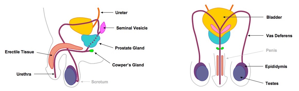
The first two functions listed above happen in the testis. There are cells in the testis that secrete the hormone testosterone into the blood from puberty onwards. Testosterone switches on secondary sexual characteristics in the male (body hair growth, muscle development, change in pitch of voice etc.) after puberty. The main part of the testis is made up of very long coiled tubules called seminiferous tubules in which the sperm cells are made.
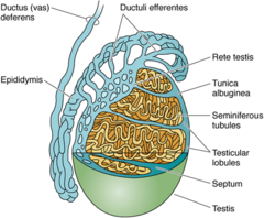
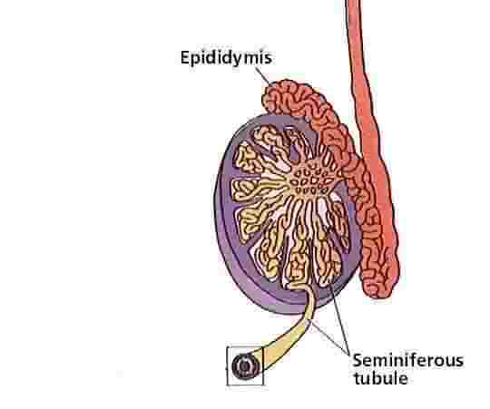
Humans have over a hundred meters of seminiferous tubules in total in both testes and this allows sperm cells to made at a very fast rate. Even though it takes around 75 days to make an individual sperm cell, the testes make them at a rate of around 85 million sperm cells per day. The epididymis is found next to the testis in the scrotum and is a coiled tube in which sperm cells continue to develop and mature. Sperm are stored here too in readiness for ejaculation.
Everyone knows that in humans the testes are found outside the body cavity in order to keep them cool. Sperm production happens at a maximal rate 3 degrees below core body temperature and having testes outside the body keeps them at this temperature.
The vas deferens is a tube lined with smooth muscle that carries sperm cells away from the testis for ejaculation. As you can see it loops around the back of the bladder, before joining up with the urethra just below the bladder. The urethra is the tube that carries urine away from the bladder but can also carry semen once the vas deferens has joined with it.
There are three accessory glands in the male system (prostate gland, seminal vesicle and the Cowpers‘ glands) These glands produce the fluid that when mixed with the sperm cells is called semen. Semen contains a sugar fructose to provide energy for the sperm cells to swim. It is slightly alkaline to neutralise the acidity in the vagina and also contains mucus to make the fluid easy to move along the tubes.
The sperm cells only acquire the ability to swim when in the epididymis and only become totally mature and able to fertilise the egg right next to the egg cell in the female tract.
The penis is an organ that contains erectile tissue that can fill with blood to allow the penis to fit into the female vagina for ejaculation.

Chromosomes video – a short PMG summary of some important ideas for GCSE
This is my first attempt at making a short summary video as a follow up to lessons. I apologise for the poor sound in places – I hope the content makes sense. Please tweet or leave a reply with any questions.
Chromosomes: Grade 9 Understanding for IGCSE Biology 3.15 3.32
I hope everyone reading this blog knows the definition of a gene. It is one of the few things in the iGCSE course that it is worth learning by heart.
“A gene is a sequence of a DNA molecule that codes for a single protein“.
In human cells, every nucleus contains about 23,000 genes. Remember there is about 1.5m of DNA inside each nucleus. For most of the life-cycle of the cell, this DNA is in a tangled web called chromatin. Chromatin is DNA molecules loosely associated with some scaffolding proteins. The scaffolding proteins are shown in the second level down of this excellent diagram as “beads on a string”.
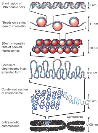
But this tangled web of DNA in chromatin poses a problem for the nucleus. For the cell to divide by mitosis, it is essential that the nucleus replicates into two identical nuclei, one for each new cell. The DNA molecules in the nucleus will make a copy of themselves by semi-conservative replication but how then can you ensure that each daughter nucleus gets exactly one copy of each DNA molecule if they are all tangled up….? This is where chromosomes come in!
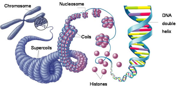
Each chromosome is a physical structure formed by supercoiling of the DNA round the scaffold proteins. The DNA coils, then folds back on itself, then coils again until each DNA molecule is so tightly coiled up that a visible chromosome appears in the nucleus. Chromosomes only become visible just before mitosis starts as for the rest of the time, the DNA is much more loosely coiled and so cannot be seen.
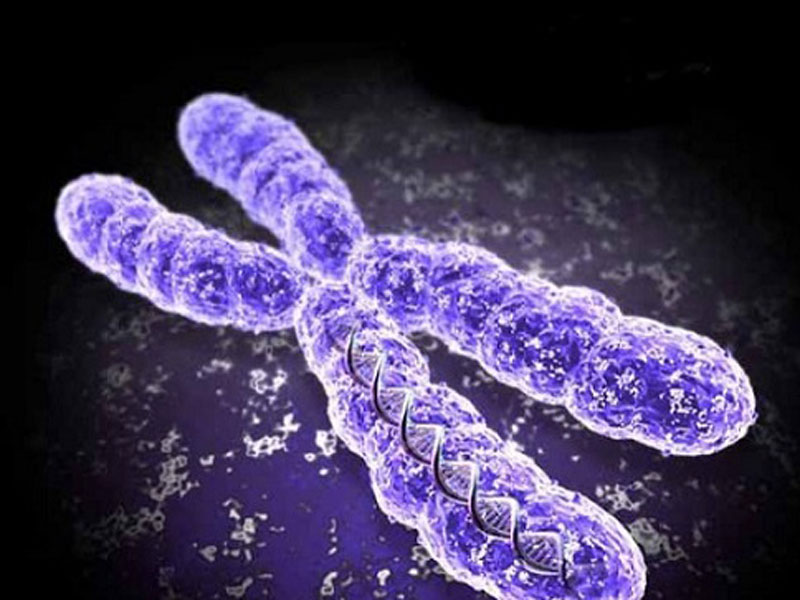
This also explains why each chromosome always looks X shaped. When chromosomes become visible the DNA has already replicated, so one chromosome is now made of two identical sister chromatids joined at a region called the centromere.

So the picture on the left shows a chromosome made as a single structure comprising one DNA molecule wrapped around the scaffold proteins. Then DNA replication occurs (in the S phase of the cell cycle) and now each chromosome is made of two identical chromatids joined at the centromere. Then the two chromatids are separated in mitosis and the chromosome returns to the structure it had at the start.
How many chromosomes are there in human cells?
The key idea here is that chromosomes are found in pairs in all body cells apart from gametes, These pairs of chromosomes (called homologous pairs) have exactly the same genes in the same locations on the chromosome. They are inherited one from each parent so one member of each pair will come from your father, one from your mother.
Different species have different numbers of pairs of chromosomes. For humans you should know that we have 23 pairs of chromosomes in the nucleus of every body cell (making a total of 46). Cells with chromosomes found in pairs are called diploid cells. Every cell in the body is diploid apart from the gametes. Gametes only have one member of each homologous pair and are called haploid cells.
Which of the following cells are diploid, which are haploid?
- Zygote
- Skin cell
- Sperm cell
- Liver cell
- Pollen grain
- Egg cell
If you are not sure, ask me by leaving a comment below….
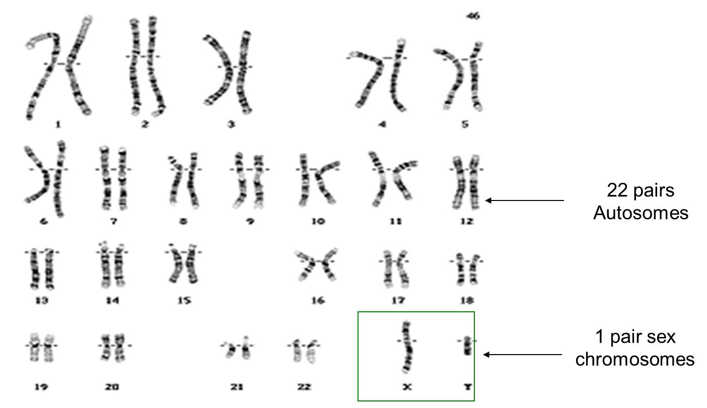
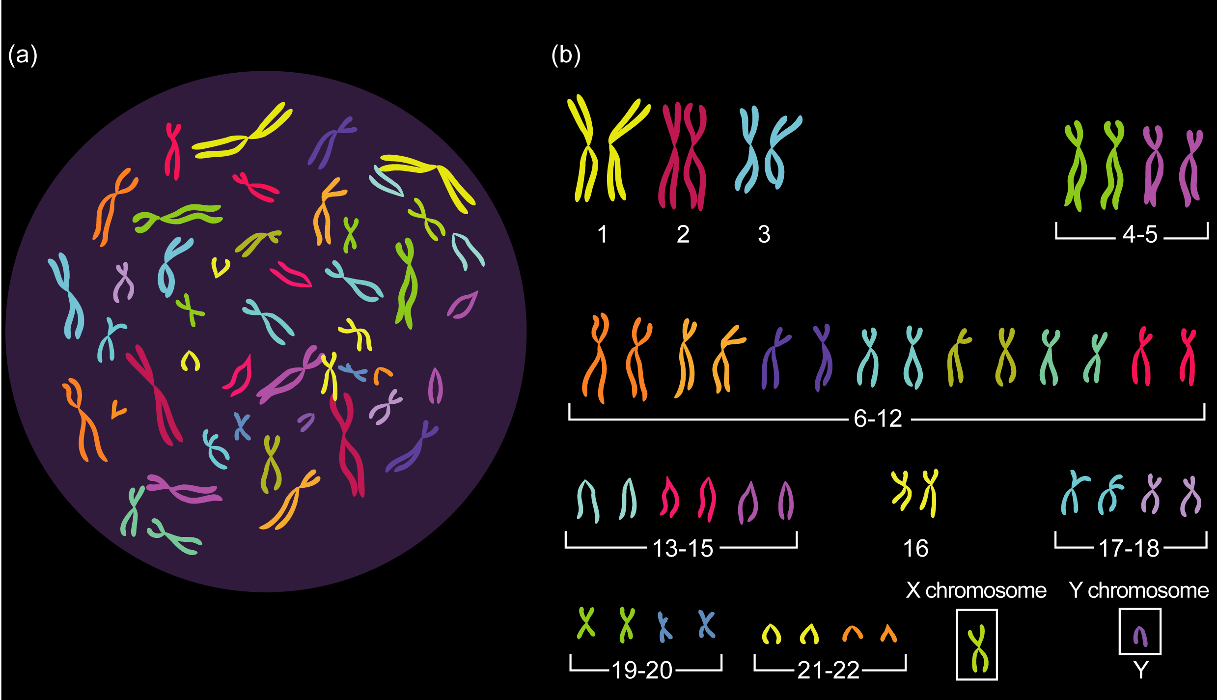
Finally for this post, chromosomes determine the sex of a human. You can see in the picture above that the 23 pairs of chromosomes can be divided into pairs 1 to 22 – these are called autosomes and play no role in determining your sex. But the 23rd pair of chromosomes are called the sex chromosomes. Males have one large X chromosome and one tiny Y chromosome as their 23rd pair whereas females have two large X chromosomes.
Gametes are haploid so only have one member of each pair. So when a man makes sperm cells (by meiosis) 50% of his sperm cells will contain his X chromosome, 50% his Y chromosome. A woman’s egg cell will always contain one X chromosome. (Why is this?) So I hope you can see that at the moment of fertilisation, the babies sex is determined depending on whether it is a Y-containing sperm cell that happens to fertilise the egg or an X-chromosome containing sperm… If the former, the baby is male, if the latter female.
I might explain this more fully in a post some other time….
Final thing for this post. If you have got to the end of this and understand everything in the text above, you are in a tiny minority of school students. Well done! This is a tricky topic and if you really understand chromosomes, you stand a chance of understanding cell division and genetics.
“Cloning a Mammoth” 5.19B 5.20B
This is a great 3 min video from the new “Hay Levels” Youtube channel. Interesting links to IGCSE Biology sections on animal cloning.
DNA is Unstable! Luckily your Cells can handle that.
I like this site – well written summary of the work done to win the most recent Nobel prize on how DNA repairs.
Another Nobel Prize story?! DAMN RIGHT! This time it’s the prize for chemistry, and Tomas Lindahl, Paul Modrich, and Aziz Sancar will collectively bask in the glory for their outstanding work in studying the mechanisms of DNA repair. Given the billions of cell divisions that have occurred in your body between conception and you today, the DNA that is copied each time remains surprisingly similar to the original that was created in the fertilized egg that you once were. Why is that strange? Well from a chemical perspective that should be impossible, with all chemical processes being subject to random errors from time to time. Along with that, DNA is subjected to damaging radiation and highly reactive substances on a daily basis. This should have led to chemical chaos long before you even became a foetus! Now, I would hope that’s not the case for you, so how do our cells prevent this…
View original post 1,695 more words
