Category: Section 2: Structures and Functions in Living Organisms

Cardiac cycle and the Human Heart: Grade 9 Understanding for IGCSE Biology 2.65 2.66
The human heart is an organ found in the middle of the thorax. It is made from a specialised type of muscle called cardiac muscle and acts as a pump to push blood around the two circulatory systems. In fact it is better to think of the heart as two separate pumps, one for the pulmonary circulatory system to the lungs and one for the systemic system that supplies blood to all the other body organs.
The diagram above shows in a simplified way this double circulatory system. The lungs are supplied with deoxygenated blood direct from the heart in the pulmonary artery but when the blood has passed through the capillaries in the lungs, it travels back to the heart in the pulmonary vein before being pumped in the systemic system around the body. Although cardiac muscle looks similar to the muscle that attaches to bones and moves the skeleton, it differs functionally in one important way. Cardiac muscle is myogenic: this means that the muscle fibres will contract without the need for a nerve impulse from the brain to initiate the contraction. Incidentally this is why a heart transplant is a possible surgical procedure. A transplanted heart will beat happily in the new body even though all the nerves going to the heart will have been cut in the surgery. You cannot have a biceps transplant at the moment because the transplanted biceps muscle would not do anything in the new patient. For the transplanted biceps to contract, the millions of individual neurones going to it would need linking up individually and this is not possible.

This is a simplified diagram showing the structure of the heart. You can see that the left and right sides of the heart are completely separate from each other. This is essential because the right side of the heart contains deoxygenated blood and the left side oxygenated blood. There are four chambers in the heart: two small atria at the top, and two larger ventricles at the bottom. The atria collect blood from the veins, the ventricles pump blood out of the heart into arteries.
There are four sets of valves in the heart: the easiest way to remember where they are is to think that blood has to pass through a set of valves as it leaves each chamber. Valves allow blood to flow through them in one direction only. The AV valves stop blood going back from the ventricle into the atrium when the ventricle contracts, the aortic and pulmonary valves (not labelled on the diagram above for some reason….) prevent blood falling back into the ventricles in between heart beats.
You also need to know the four main blood vessels that are attached to the heart. The vena cava is the largest vein in the body and carries deoxygenated blood from the organs of the body into the right atrium. The pulmonary artery comes out of the right ventricle and pumps this deoxygenated blood to the lungs. Oxygenated blood returns from the lungs to the left atrium in the pulmonary veins, passes into the left ventricle and is then pumped out of the heart in the aorta, the biggest artery in the body.

NB I strongly suggest you do not use the terms bicuspid, mitral or tricuspid valve to label the valves between the atria and ventricles… In an exam it is easy to get these similar words muddled, so I would always call the valve between the left atrium and left ventricle the left atrio-ventricular valve (never bicuspid or mitral valve) and I would always call the valve between the right atrium and right ventricle the right atrio-ventricular valve (never tricuspid). I know many of you will ignore this advice but it’s good to get it off my chest……
I’ve just spent 45 minutes trying to find a good video on heart structure to put into the post but without success…. If anyone knows a really good YouTube clip (must be under 5 minutes) please add a link as a comment at the foot of this post.
Events of the Cardiac Cycle
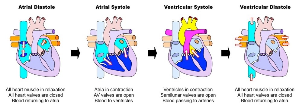
This is a topic that requires some simplification as it is easy to get confused. The Cardiac Cycle is simply a word for the sequence of events that happen in a heart beat. At the simplest level, the cardiac cycle consists of three phases:
1. Diastole. During this stage the cardiac muscle is relaxed (the heart is between beats) and blood can enter the atria and then fall into the ventricles through the open AV valves.
2. Atrial Systole. This stage in when the cardiac muscle in the atria contract, increasing the atrial pressure and pushing blood down into the ventricles. There is a small region in the wall of the right atrium called the sino-atrial node (or pacemaker) which initiates each heart beat.
3. Ventricular Systole. After a short delay, the cardiac muscle in the ventricles contracts. This increases the blood pressure in the ventricle which in turn causes the AV valve to close, and the aortic or pulmonary semilunar valves to open. As these valves at the exit of the ventricle open, blood gets pushed into the arteries and out of the heart.
Look at the diagram above and make sure you understand the meaning of these three terms.
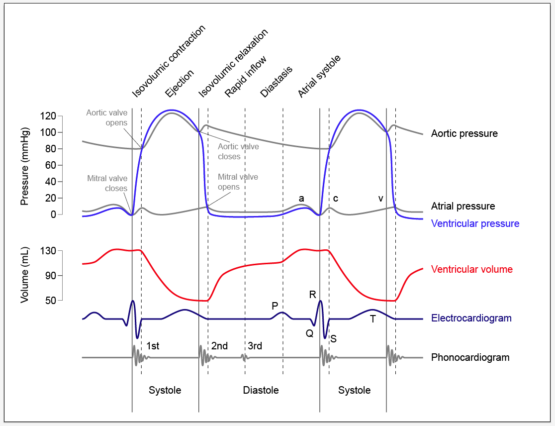
The diagram below is much more complex and perhaps you should not worry too much about it….. The important bit is to understand when the valves in the heart open and when they close. It is quite simple really although you would be amazed how confused people can get….
The opening and closing of a valve is not “controlled” in any meaningful way in the heart. The valve has a structure that will only allow it to open in one direction. Let’s consider the AV valves. These can open to allow blood to pass from the atria into the ventricles during atrial systole but will close during ventricular systole to stop the blood flowing back where it came from.
The AV valve will be open whenever the blood pressure in the atria is greater than in the ventricle.
The AV valve will be closed whenever the blood pressure in the ventricle is greater than in the atrium.
Look at the graph of pressure changes below. You can see the mitral valve (I hate that name) is closed as soon as the ventricular pressure exceeds the atrial pressure and it opens again during ventricular diastole as the pressure in the ventricle drops.

Blood part 2 White Blood Cells – Grade 9 Understanding for GCSE Biology 2.59 2.62 2.63B
The previous post looked at the structure and function of red blood cells and plasma. Now it is time to turn our attention to the rather more complex topic of white blood cells….. This is a topic in which the complexity can put people off but I am deliberately going to keep things simple (I hope!). If you are thinking about revision for GCSE, don’t worry about anything more complicated than in this post.

There are many types of white blood cell found in blood. But let’s keep things simple…. You need to understand the role of lymphocytes and phagocytes in defending the body against pathogens.
A pathogen is defined as “a microorganism that can cause a disease” and pathogens may be bacteria, viruses, protistans or fungi. Can you give me an example of an infectious disease caused by each class of pathogen?
The structure of these two classes of white blood cell is important. The commonest phagocytes in blood are called neutrophils and they are easily recognised by their irregular shaped nucleus and cytoplasm packed full of granules. Lymphocytes are much smaller white cells and are identifiable by their clear cytoplasm and large spherical nucleus that takes up 90% of the volume of the cell.


So now we should look at how these two types of white blood cells defend the body against pathogens. Remember that the account on this post is an over-simplification of what is in reality an extremely complex process.
Let’s start with a phagocyte. These large cells are able to engulf invading pathogens in the blood and tissue fluid by a process called phagocytosis.

The phagocyte pushes out projections of its cytoplasm around the clump of bacteria. These projections are called pseudopodia and when they meet, the cell membrane of the phagocyte fuses together leaving the bacteria enclosed in a tiny membrane packet called a vesicle inside the cytoplasm. The phagocyte then fuses other vesicles that contain powerful digestive enzymes with the vesicle with the bacteria in, leading to the death and destruction of the bacteria. Simple.
The problem for phagocytes is this: how do they know what to engulf and destroy? This is where lymphocytes come in. One class of lymphocyte is able to secrete small soluble proteins called antibodies into the blood. Antibodies are specific to a particular surface marker on the invading pathogen and bind to it because the shape of the antibody and the shape of the surface marker are complimentary.

Now people always get confused between antibodies (the small soluble Y-shaped proteins secreted by lymphocytes) and antigens (the surface markers on the invading pathogen). Make sure you are completely clear on the difference in meaning of these two words….
 This diagram shows antibodies (green) binding to surface markers (antigens) on a bacterial cell.
Antibodies produced by lymphocytes will coat the invading pathogen by binding to antigens on its surface. One effect of this is that phagocytes are stimulated to engulf the antibody-coated organism.
There are many different types of lymphocyte and not all can produce antibodies. Another important function of lymphocytes is to kill your own body cells when they are corrupted, either by the presence of a virus or by becoming cancerous.
Finally, can I draw your attention to two previous posts linked to this one. The first is on the role of platelets in blood clotting, the second on the difficult topic of immunity and how lymphocytes are responsible for giving you lifelong protection against certain infectious diseases.
https://pmgbiology.wordpress.com/2014/04/07/immunity-a-understanding-for-biology-igcse/
As always, please ask me questions either via the comment section below the post or with a tweet…. I will do my best to respond to any questions from anyone who is bothered to read my posts!
Blood part 1 Plasma and RBCs: Grade 9 Understanding for IGCSE Biology 2.59, 2.60, 2.61
Blood is a tissue in the body that plays a variety of roles in transport and in defending the body against disease. It is an unusual tissue since it is a liquid, with many different kinds of cells suspended in a watery solution called plasma. 
Plasma makes up 55% of the volume of blood and is a solution of many different chemicals in water. For example, the plasma contains dissolved glucose, amino acids and other products of digestion from the intestines. It also transports the waste molecule urea from the liver where it is made to the kidney where it is excreted. Blood plasma contains dissolved carbon dioxide, mostly in the form of hydrogencarbonate ions. Many hormones (for example testosterone, ADH, adrenalin) are transported in the blood plasma and because the plasma is mostly water, it provides a good way of moving heat around the body from respiring muscles to the skin where it can be lost.
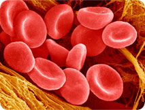
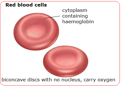
The most common cell in blood are the red blood cells (or erythrocytes). These tiny cells are adapted for the transport of oxygen. Each red blood cell contains around 270 million molecules of a transport protein, haemoglobin. Each molecule of haemoglobin can bind up to four molecules of oxygen in the lungs and then unload the oxygen when the red blood cell passes through a capillary in an actively respiring tissue.

(Don’t worry too much about the structure of the protein – this is A level stuff really…. Just remember haemoglobin is a transport protein for oxygen found in red blood cells)
As well as being packed full of haemoglobin molecules, red blood cells have other adaptations for transporting oxygen. Red blood cells lose their nucleus during their development as this allows more haemoglobin to be packed into each cell. Having no nucleus means the red blood cell cannot divide nor repair damage to its structure. This is why each red blood cell only lives for 100-120 days in the body.
Red blood cells have a characteristic shape. It is called a biconcave disc and they have an especially flexible shape. Remember that a capillary is actually smaller in diameter than a red blood cell, so the cells have to squeeze through capillaries in single file…..

Blood vessels – Grade 9 Understanding for IGCSE Biology 2.68
In this post, I will look at the structure and function of the three main types of blood vessel in the human circulatory system. Although this is not the most difficult topic, there are a few things that can catch out even A* GCSE students in the heat of an exam.
Arteries are the blood vessels that take blood away from the heart. Because the blood is coming straight from the ventricles of the heart, it will be at a high blood pressure and will flow in pulses. This means that arteries need a thick wall to withstand this high blood pressure. All arteries apart from one carry oxygenated blood. Can you remember which artery is the exception to this rule?

The artery wall has a narrow lumen (the space where the blood flows) as this helps to maintain the high blood pressure within. There are also many elastic fibres in the middle tissue (tunica media) of the artery wall. This elastic tissue is important because the blood flows in pulses. The artery wall needs to stretch as the pulse of blood passes and the elastic recoil of the wall helps to push the blood along in between heart beats.
The tunica media also contains a lot of smooth muscle. Why do arteries need muscle in their walls? When this muscle contracts it narrows the lumen of the vessel. This will increase the blood pressure and so one reason for muscle in arteries is to regulate the blood pressure. But there is something more… Arteries carry blood into the organs of the body and the pattern of blood flow to different organs can vary depending on the conditions. For example, when you are running, you need more blood to go to your skeletal muscles (to carry oxygen for respiration and to remove heat and carbon dioxide) and less to go to the digestive system. This is brought about by the smooth muscle in the artery taking blood to the intestines and stomach contracting so that less blood can flow through the vessel. The smooth muscle in the arteries in the exercising muscles will relax so that more blood can pass. This shift in the pattern of blood flow is the second key significance of arteries having lots of muscle in the walls.

Veins have the same tissues in their walls as arteries but they are much thinner. The blood is flowing at a much lower pressure in veins as all the pressure from the heart has been lost in the extensive capillary beds in the tissues. Veins return blood to the heart and all bar one (the pulmonary vein) contain deoxygenated blood. As there is low blood pressure in veins, this can cause problems moving blood back to the heart especially when against gravity. Veins contain valves which only allow blood through in one direction thus preventing the blood falling back. The thin walls of veins also mean that they can be compressed by the action of skeletal muscles. When the muscles that move the skeleton contract, they can squeeze on veins and help to return blood to the heart.

Capillaries are the smallest of the three types of blood vessel. They are found in the tissues throughout the body and are beautifully adapted to ensure the exchange of materials between the cells of the body and the blood. The lumen of a capillary is less than the width of a red blood cell and so red blood cells pass through capillaries in single file and only be squeezing along. This ensures the speed of blood flow in capillaries is very very slow.
The lining of a capillary is made up of a single layer of cells called the endothelium. Arteries and Veins have an endothelium too but in the capillary the endothelial cells have gaps between them called pores. This allows the fluid component of blood and various white blood cells to leak out of capillaries to form tissue fluid. This leaky nature of capillaries is very important as it provides the fluid that bathes the tissues of the body.

Enzymes: Grade 9 Understanding for IGCSE Biology 2.10 2.11 2.13
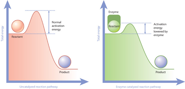
Enzymes are biological catalysts. This means they are able to increase the rate of a chemical reaction but are not used up in the reaction. Without enzymes the reactions of metabolism would all happen too slowly for life to exist – enzymes can speed up the rate of reaction by many millions of times….
Enzymes work as catalysts by lowering the activation energy needed for the reaction to occur. Activation energy is the term for the extra energy needed to be given to the reactants to break bonds within them to allow the product molecules to be formed. Enzymes provide an alternate reaction pathway that has a lower activation energy. This means that under any conditions a higher proportion of the reactant molecules will have sufficient energy to overcome the activation energy barrier and so more reactants will be turned into products.
How do enzymes lower the activation energy?
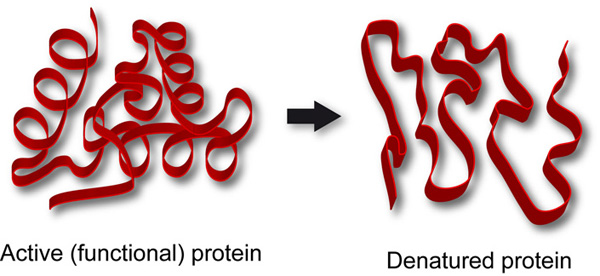
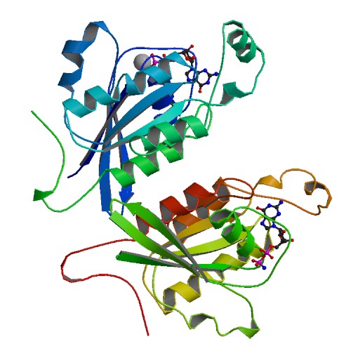
Enzymes are all large globular molecules, almost always made of protein. They have a specific three-dimensional shape that includes a region called the active site which has a shape that allows the reactant molecules (called substrates) to bind. When the enzyme binds to the substrate, it forms an enzyme-substrate complex.

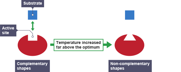
This theory of how enzymes might work is called the Lock and Key theory. The active site acts like a lock as it has a shape that is complementary to the shape of the substrate (the key). Lock and Key theory explains an important property of enzymes which is that they are specific. Each enzyme can only catalyse one reaction since only a substrate molecule with a specific shape can bind to the active site.
When the substrate is bound to the active site forming an enzyme-substrate complex, the enzyme introduces a strain on some of the bonds in the substrate, making a reaction more likely. The active site might provide a microenvironment that is exactly the right condition for the reaction, thus lowering the activation energy.

Key idea: enzymes catalyse almost all the chemical reactions that happen in organisms. It is easy to imagine that enzymes only catalyse reactions like the one in the picture above in which a molecule is being broken down into smaller molecules. But enzymes catalyse oxidation reactions, condensation reactions in which big molecules are built up from smaller ones, phosphorylation reactions (sticking phosphate groups onto molecules) and so on and so on. So don’t describe enzymes as being involved in breaking things down: some do of course but the vast majority work inside cells to catalyse a whole variety of reactions in metabolism.
Rates of enzyme-catalysed reactions can be affected by Temperature
The temperature of the reaction has a significant effect on the rate of reaction. Look at the following graphs:

The pattern of this graph is characteristic of an enzyme catalysed reaction. At low temperatures the rate of reaction is low. This is because few enzyme-substrate complexes are formed per second as the enzyme and substrate molecules are moving around so slowly that they rarely collide. At temperatures above the optimum, the enzymes and substrate molecules will be moving very fast and so will be colliding all the time. So why is the rate so low? Well that is because high temperatures cause enzymes to denature. Remember enzymes are made of protein and proteins can have their 3D shape changed by high temperatures. If an enzymes’ 3D shape changes, the active site will change shape and if this happens the substrate cannot bind.
Rates of enzyme-catalysed reaction are also affected by pH
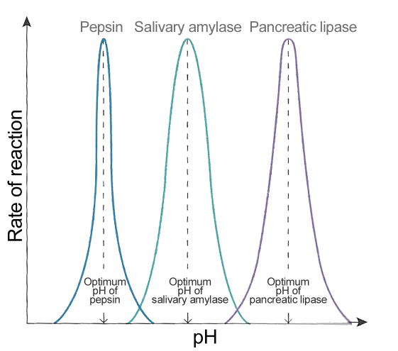
Enzymes tend to work in a very narrow band of pH values. pH is a measure of the acidity/alkalinity of a solution and most enzymes require an optimum pH to function well. The rate of reaction drops very rapidly on either side of the optimum pH simply because extremes of pH will denature enzymes. The acid or alkaline environment can break the bonds that hold the enzyme in its specific 3D shape. An enzyme with a changed shape cannot function as a catalyst if the substrate cannot bind to the active site and so the rate falls away rapidly either side of the optimum.

Factors affecting rates of Photosynthesis (part 2): Grade 9 Understanding for IGCSE Biology 2.20 2.23
In the previous post on photosynthesis, you revised how there were four environmental factors that can affect rates of photosynthesis in a plant:
- light intensity
- light wavelength
- temperature
- carbon dioxide concentration
This post will explain the results from experiments with Elodea in which one factor is altered (the independent variable) and the other three are kept exactly the same (control variables)
Light intensity

The independent variable (light intensity) is on the x axis and the dependent variable (number of bubbles per minute) is on the y axis.
How do we explain the pattern in this graph?
As the light intensity increases the rate of photosynthesis increases. This is because a higher light intensity gives more energy to the chloroplasts and so more reactions can happen per second and the rate goes up. But beyond the orange dot on the graph, the increases in rate slows down until at around 12 units of light, adding more light has no effect on the rate. At these high light intensities some other factor is now the limiting factor as opposed to light intensity. The limiting factor remember is the factor in the shortest supply. So perhaps above 12 units of light photosynthesis is limited by the concentration of carbon dioxide. The only way to find the limiting factor is to repeat the experiment with more carbon dioxide and see whether the rate is higher above 12 units.
Light wavelength
Although this graph is not perfect, it does show how the rate of photosynthesis varies at different light wavelength.

Rates of photosynthesis peak in the blue-violet and red parts of the visible spectrum with a much lower rate in green light. The reason for this is that chlorophyll pigments do not absorb green light well.
Carbon Dioxide concentration

The pattern is similar to the light intensity relationship. When carbon dioxide concentrations are low, it is the limiting factor for photosynthesis and so increasing the concentration will increase the rate. As the graph levels off, some other factor is now the limiting factor – perhaps light intensity or temperature.
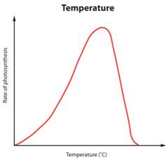
Temperature
Temperature is a factor that affects photosynthesis because of enzymes. Many reactions in photosynthesis are catalysed by enzymes and enzymes all have an optimum temperature.
This pattern is not explained by limiting factors. At low temperatures the rate is low because the enzymes and the substrate molecules are moving really slowly. This means there are few collisions between the substrate and the active site of the enzyme. As temperature increases, the rate increases as there are more collisions and more enzyme-substrate complexes are formed per second. But high temperatures denature enzymes: the bonds that hold the enzyme in its precious 3-D shape are broken and the enzyme molecule unravels. So the active site may either change shape or may be lost as a catalyst. This slows the rate down to an extremely low rate.

Factors affecting rates of Photosynthesis: Grade 9 Understanding for IGCSE Biology (part 1) 2.18 2.19
Photosynthesis is the process occurring in plants in which sunlight is trapped by chlorophyll pigments and used to power the chemical reactions involved in making food molecules such as carbohydrates from carbon dioxide and water. Oxygen is released as a waste product of these reactions.
(I can’t write a chemical equation as I can’t find a way of writing subscript in WordPress….. Can anyone help?)

In the equation above, the carbohydrate produced is glucose, a six carbon sugar.
The reactions of photosynthesis happen in specialised mesophyll tissue in the leaf of the plant (see previous post) Inside the palisade and spongy mesophyll cells there are thousands of tiny organelles called chloroplasts in which the reactions of photosynthesis occur.
So what environmental factors could be altered to vary the rate of photosynthesis in a plant?
Light Intensity – light provides the energy for photosynthesis and so the higher the intensity of light, the more energy the chloroplasts receive to make carbohydrates.
Light wavelength – chlorophyll pigments absorb the blue-violet and red parts of the spectrum well but cannot absorb green light.
Carbon Dioxide concentration – this is a reactant for photosynthesis so increasing the concentration makes a collision between the reactant molecule and the enzyme inside the chloroplast that bind it more likely, so the rate will go up.
Temperature – many reactions in photosynthesis are catalysed by enzymes and enzymes are very affected by temperature: too low temperatures and the enzymes and substrate molecules move very slowly and so there are few collisions, too high temperatures and the enzymes change shape (denature) so the substrate molecules cannot fit into the active site.
NB – water availability is never a factor that can alter rates of photosynthesis even though it is a reactant molecule. This might seem unusual until one remembers that plants that are dehydrating will close the stomata in their leaves to minimise transpiration. Closed stomata mean that carbon dioxide cannot get into the air spaces in the leaf so this is ultimately what limits photosynthesis in a dehydrated plant.
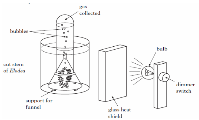
The experimental set up above is the best way to measure rates of photosynthesis and so investigate the effect of any of the four factors listed above. Light intensity can be varied either with a dimmer switch as above or by altering the distance between the lamp and the plant. The heat shield is transparent to let light through but will absorb the heat from the bulb ensuring the temperature of the water stays constant. Carbon dioxide concentration can be altered by dissolving different masses of sodium hydrogen carbonate in the water. The wavelength of light will stay constant so long as the build remains the same.
How to measure rates of photosynthesis in this set up?
Well you could collect the gas produced over a long period of time and measure its volume with a gas syringe. This might sound more accurate than counting bubbles but in fact it is a less reliable way as you would have to leave the set up for a long time and variables might change. So it is fine to assume that the bubbles produced are oxygen and that every bubble is the same volume: if you do this, the rate of production of bubbles is directly proportional to the rate of photosynthesis in the Elodea plant.

Diffusion, Active Transport and Osmosis: Grade 9 Understanding for IGCSE Biology 2.15 2.16
This post is going to describe some of the ways molecules can cross the cell membrane. (For Eton students revising for Trials, diffusion and active transport are found in the F block syllabus, osmosis comes in E Block)
Diffusion is the simplest to understand. Diffusion does not even need a cell membrane to occur. In the example below the dye molecules will move randomly in the solution. As the dye starts in one place, these random movements will mean that slowly spread out until an equilibrium is reached. This movement of the dye from the region of high concentration to the low concentration is called diffusion.

When considering diffusion into a cell, if the cell membrane is permeable to a particular molecule then the random movements of the molecule will mean that there will be a net (overall) movement from the higher concentration to the lower concentration down the concentration gradient.

Key Points about diffusion:
- Always happens down a concentration gradient (from a high concentration to a lower one)
- Never requires any energy from the cell – it is a passive process
Active Transport is a process that will move molecules into a cell against the concentration gradient – i.e. from a low concentration to a high concentration. This “pumping” of the molecules against the gradient requires energy from the cell and of course this energy comes from respiration.

You can see from the diagram above that active transport is working against the concentration gradient, is using energy from inside the cell (actually a molecule made in mitochondria in respiration called ATP) and that a specific transport protein is involved in the cell membrane. This protein will have a binding-site that is specific for a particular molecule and the solute molecule to be transported will collide with the transport protein due to random movement. Energy from the cell can cause the transport protein to change shape such that the solute is released on the other side of the membrane.
Can you think of another area of the iGCSE syllabus which features collisions between a specific binding-site on a protein and a certain other molecule? Linking ideas is a key characteristic of the A* Biologist!

Osmosis is the hardest of these processes to understand properly, especially as an iGCSE student when you are often told an over-simplified account that does not make sense…. Let’s try to simplify it in a way that does make sense.
Firstly it is only water molecules that can move by osmosis into and out of cells – never anything else. Indeed osmosis is the only way water can cross a membrane – it never moves by diffusion or active transport.
Osmosis is a passive process – it never needs any energy from the cell’s respiration and the only energy involved is the kinetic energy of the water molecules.
Osmosis can only occur through a partially permeable membrane. All cell membranes are partially permeable and this means they let small molecule like water through but prevent the diffusion of the larger solute molecules.

The water molecules on both sides of the membrane in the diagram above will be moving around randomly. They will occasionally hit one of the pores in the membrane and so pass across the membrane. This movement will be happening from left to right and from right to left.
But….
The presence of the sucrose (solute) in the solution on the right means that some of the water molecules on that side of the membrane are less able to move. This is because they are temporarily attracted to the solute molecules by weak hydrogen bonds. So their kinetic energy is reduced and this makes them less likely to randomly collide with the pores in the membrane. The presence of the solute on the right means that water molecules on the left on average are more likely to collide with the membrane than the water molecules on the right and this leads to an overall movement from left to right. This net movement of water molecules from the dilute solution to the more concentrated solution through the partially permeable membrane is called osmosis.

This diagram has the two solutions reversed so in which direction will osmosis happen here? Thats right from right to left. You can see the hydrogen bonds attracting water molecules to the solute – these are the ones that lower their kinetic energy overall.
You might even have been taught about osmosis with reference to the water potential of a solution. The water potential of a solution is just a measure of how much kinetic energy the water molecules in a solution possess. So a dilute solution will have a high water potential, a concentrated solution (with lots of dissolved solute) a lower water potential.
Osmosis is the
- net movement of water
- through a partially permeable membrane
- from a solution with a high water potential (a dilute solution) to a solution with a lower water potential (a concentrated solution)
Biological examples
Diffusion
- Oxygen diffuses from the air in the alveolus into the blood
- Carbon Dioxide diffuses from the air spaces in the leaf into the palisade mesophyll cells of the leaf
- Glucose diffuses from the blood into an actively-respiring muscle
Active Transport
- Nitrates are pumped from the soil into root hair cells by active transport
- In the kidney, glucose and other useful molecules are pumped from the nephron back into the blood by active transport.
- In nerve cells, sodium and potassium ions are pumped across the cell membrane to set up the gradients needed for a nerve impulse
Osmosis
- Water enters root hair cells from the soil by osmosis
- In the kidney, water is reabsorbed from the nephron by osmosis.
- In the large intestine, water is reabsorbed from the colon back into the blood by osmosis
There are many many more examples of each process, but this should be enough to be going on with…….
Leaf structure and Adaptations for Photosynthesis: Grade 9 Understanding for IGCSE Biology 2.21
The leaf is the organ in a plant specially adapted for photosynthesis. You need to understand the structure of the tissues in a leaf together with their functions.

Upper Epidermis: this is the tissue on the upper surface of the leaf. It produces a waxy layer, called the cuticle, which is not made of cells but is a waterproof barrier to prevent excessive evaporation through the hot upper surface of the leaf. The upper epidermis cells have no chloroplasts so light passes through them easily.
Palisade Mesophyll: this tissue is where 80% of the photosynthesis takes place in the leaf. The palisade cells have many chloroplasts in their cytoplasm and the box-like shape and arrangement of these cells ensures they are packed tightly together.
Spongy Mesophyll: this tissue contains large air spaces which are linked to the atmosphere outside the leaf through microscopic pores called stomata on the lower surface. Spongy mesophyll cells also contain chloroplasts and photosynthesis occurs here too. The air spaces reduce the distance carbon dioxide has to diffuse to get into the mesophyll cells and the fact that these cells have fairly thin cell walls which are coated with a film of water together means that gas exchange between air space and mesophyll is speeded up.
Lower Epidermis is the most dull tissue in the leaf. The only interesting thing about it is that it contains specialised cells called guard cells which enclose a pore called a stoma. Carbon dioxide can diffuse into the leaf through the stomata when they are open (usually at day time) and water evaporates out of the stomata in a process called transpiration.
Adaptations of a Leaf for Photosynthesis
- Large Surface Area – to maximise light harvesting
- Thin – to reduce distance for carbon dioxide to diffuse through the leaf and to ensure light penetrates into the middle of the leaf
- Air Spaces – to reduce distance for carbon dioxide to diffuse and to increase the surface area of the gas exchange surface inside the leaf
- Stomata – pores to allow carbon dioxide to diffuse into the leaf and water to evaporate out (transpiration)
- Presence of Veins – veins contain xylem tissue (carries water and minerals to the leaf from the roots) and phloem (transports sugars and amino acids away from the leaf)
- Chloroplasts – mesophyll cells and guard cells contain many chloroplasts. These organelles contain the light harvesting pigment chlorophyll and are where all the reactions of photosynthesis occur

Biological Molecules: Grade 9 Understanding for IGCSE Biology 2.7 2.8
You will have studied the Biological Molecules section in some detail I would imagine, perhaps in more detail than is absolutely required for the specification. This post is meant to help you focus your understanding onto those points that are most likely to be tested in iGCSE questions. Here goes…
You do need to understand some chemistry for this topic to make sense. In particular you need to understand what is meant by the following terms:
- atom
- molecule
- element
- compound
My personal definitions would be as follows:
Atom: the smallest particle that retains the chemical properties of the element – a structure made up of protons, neutrons and electrons
Molecule: a particle made of two or more atoms chemically bonded together – may contain just one type of atom or several
Element: a substance in which all the atoms are the same
Compound: a substance containing more than one type of element
Back to safer ground…..
Living organisms are made from a fairly small group of molecules. The commonest molecule in every organism is water and in humans water makes up about 70% of the mass. But if you were to remove all water, leaving behind just the dry mass, the most common molecules could be grouped into proteins, lipids, carbohydrates and nucleic acids (e.g DNA)
Carbohydrates contain just three elements – carbon, hydrogen and oxygen
Lipids (fats and oils) contain three elements – carbon, hydrogen and oxygen
Proteins contain four or five elements – carbon, hydrogen, oxygen, nitrogen and sometimes sulphur
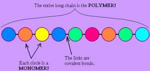
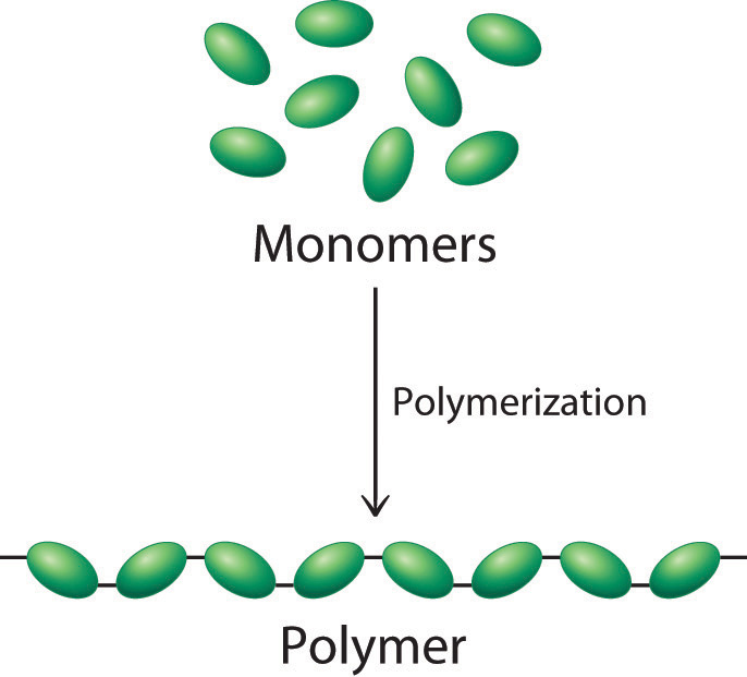
Big idea: many of the molecules that living things are made from are examples of polymers. A polymer is a large molecule made up of a long chain of repeating subunits (called monomers)
Carbohydrates are grouped into three main types:
Simple sugars like glucose or fructose – these are called monosaccharides.
Some sugars like sucrose are made of two simple sugars joined together – these are called disaccharides
Some carbohydrates are macromolecules (polymers) made of many hundreds of sugar residues joined together – these are called polysaccharides.
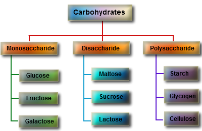
You can see from the diagram above that there are three important polysaccharides in living organisms. All three are polymers of the sugar glucose but the arrangement of the glucose residues is different. Cellulose is the main constituent of plant cell walls. Starch is a storage polysaccharide found in plants and Glycogen is a similar storage molecule found in liver and muscle tissue in animals.

Glucose is detected using a Benedict’s Test. Heat the solution with Benedict’s,reagent to 90 degrees for 5 minutes. A positive test for glucose is a brick red colour.
Starch is tested for using iodine solution (in potassium iodide) Iodine solution turns blue-black in the presence of starch.
Proteins are also polymers but this time the individual monomer is not a sugar but a molecule called an amino acid.
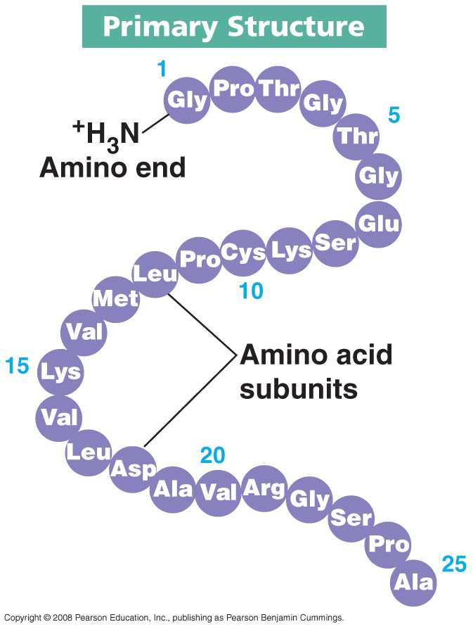
This protein is then folded up into a complex 3D shape using a whole load of weak bonds that can easily be broken at high temperatures. This is why enzymes, made of protein, denature at high temperatures.
There are 20 different amino acids that could be incorporated into a protein so there are an almost limitless variety of different proteins that can be made.
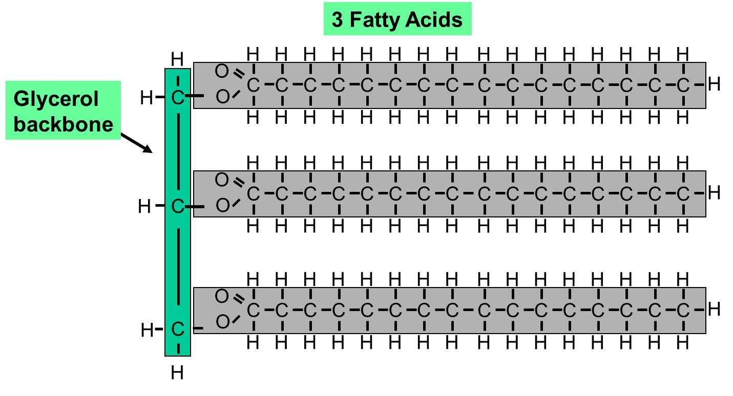
Lipids are a group of water-repelling molecules that again contain C,H and O atoms. They used to be separated into fats and oils depending in whether they are a solid (fat) or liquid (oil) at room temperature. Many lipids are a type of molecule called a triglyceride and this is made of a single molecule of glycerol attached to three fatty acid tails.