Factors affecting rates of Photosynthesis: Grade 9 Understanding for IGCSE Biology (part 1) 2.18 2.19

Photosynthesis is the process occurring in plants in which sunlight is trapped by chlorophyll pigments and used to power the chemical reactions involved in making food molecules such as carbohydrates from carbon dioxide and water.  Oxygen is released as a waste product of these reactions.

(I can’t write a chemical equation as I can’t find a way of writing subscript in WordPress….. Can anyone help?)


In the equation above, the carbohydrate produced is glucose, a six carbon sugar.

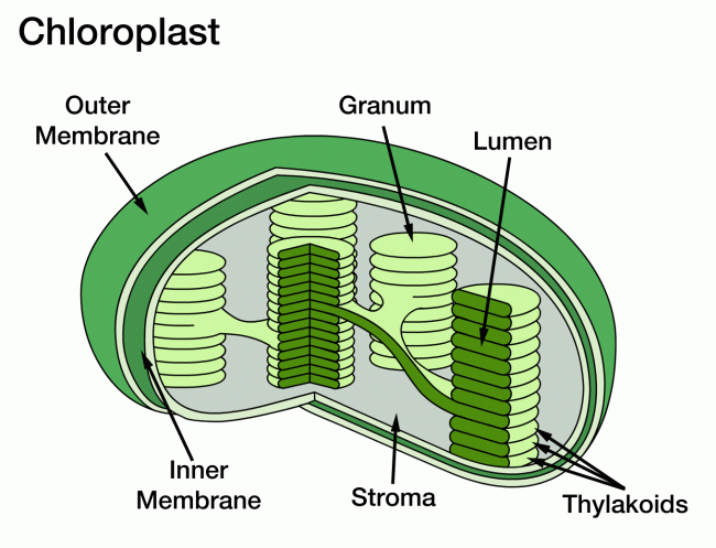
The reactions of photosynthesis happen in specialised mesophyll tissue in the leaf of the plant (see previous post)  Inside the palisade and spongy mesophyll cells there are thousands of tiny organelles called chloroplasts in which the reactions of photosynthesis occur.

Structure-of-Chloroplasts

So what environmental factors could be altered to vary the rate of photosynthesis in a plant?

Light Intensity – light provides the energy for photosynthesis and so the higher the intensity of light, the more energy the chloroplasts receive to make carbohydrates.

Light wavelength – chlorophyll pigments absorb the blue-violet and red parts of the spectrum well but cannot absorb green light.

Carbon Dioxide concentration – this is a reactant for photosynthesis so increasing the concentration makes a collision between the reactant molecule and the enzyme inside the chloroplast that bind it more likely, so the rate will go up.

Temperature – many reactions in photosynthesis are catalysed by enzymes and enzymes are very affected by temperature:  too low temperatures and the enzymes and substrate molecules move very slowly and so there are few collisions, too high temperatures and the enzymes change shape (denature) so the substrate molecules cannot fit into the active site.

NB – water availability is never a factor that can alter rates of photosynthesis even though it is a reactant molecule.  This might seem unusual until one remembers that plants that are dehydrating will close the stomata in their leaves to minimise transpiration.  Closed stomata mean that carbon dioxide cannot get into the air spaces in the leaf so this is ultimately what limits photosynthesis in a dehydrated plant.

N5_Unt_1_Sht_19a

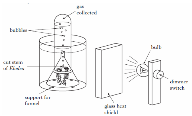
The experimental set up above is the best way to measure rates of photosynthesis and so investigate the effect of any of the four factors listed above.  Light intensity can be varied either with a dimmer switch as above or by altering the distance between the lamp and the plant.  The heat shield is transparent to let light through but will absorb the heat from the bulb ensuring the temperature of the water stays constant.  Carbon dioxide concentration can be altered by dissolving different masses of sodium hydrogen carbonate in the water.  The wavelength of light will stay constant so long as the build remains the same.

How to measure rates of photosynthesis in this set up?

Well you could collect the gas produced over a long period of time and measure its volume with a gas syringe.  This might sound more accurate than counting bubbles but in fact it is a less reliable way as you would have to leave the set up for a long time and variables might change.  So it is fine to assume that the bubbles produced are oxygen and that every bubble is the same volume: if you do this, the rate of production of bubbles is directly proportional to the rate of photosynthesis in the Elodea plant.

Diffusion, Active Transport and Osmosis: Grade 9 Understanding for IGCSE Biology 2.15 2.16

This post is going to describe some of the ways molecules can cross the cell membrane.  (For Eton students revising for Trials, diffusion and active transport are found in the F block syllabus, osmosis comes in E Block)

Diffusion is the simplest to understand.  Diffusion does not even need a cell membrane to occur.   In the example below the dye molecules will move randomly in the solution.  As the dye starts in one place, these random movements will mean that slowly spread out until an equilibrium is reached.  This movement of the dye from the region of high concentration to the low concentration is called diffusion.

diffusion_1

When considering diffusion into a cell, if the cell membrane is permeable to a particular molecule then the random movements of the molecule will mean that there will be a net (overall) movement from the higher concentration to the lower concentration down the concentration gradient.

32

Key Points about diffusion:

  • Always happens down a concentration gradient (from a high concentration to a lower one)
  • Never requires any energy from the cell – it is a passive process

Active Transport is a process that will move molecules into a cell against the concentration gradient – i.e. from a low concentration to a high concentration.  This “pumping” of the molecules against the gradient requires energy from the cell and of course this energy comes from respiration.

06-16

You can see from the diagram above that active transport is working against the concentration gradient, is using energy from inside the cell (actually a molecule made in mitochondria in respiration called ATP) and that a specific transport protein is involved in the cell membrane.  This protein will have a binding-site that is specific for a particular molecule and the solute molecule to be transported will collide with the transport protein due to random movement.  Energy from the cell can cause the transport protein to change shape such that the solute is released on the other side of the membrane.

Can you think of another area of the iGCSE syllabus which features collisions between a specific binding-site on a protein and a certain other molecule?  Linking ideas is a key characteristic of the A* Biologist!

3168392_orig

Osmosis is the hardest of these processes to understand properly, especially as an iGCSE student when you are often told an over-simplified account that does not make sense….  Let’s try to simplify it in a way that does make sense.

Firstly it is only water molecules that can move by osmosis into and out of cells – never anything else.  Indeed osmosis is the only way water can cross a membrane – it never moves by diffusion or active transport.

Osmosis is a passive process – it never needs any energy from the cell’s respiration and the only energy involved is the kinetic energy of the water molecules.

Osmosis can only occur through a partially permeable membrane.  All cell membranes are partially permeable and this means they let small molecule like water through but prevent the diffusion of the larger solute molecules.

osmosis_1

The water molecules on both sides of the membrane in the diagram above will be moving around randomly.  They will occasionally hit one of the pores in the membrane and so pass across the membrane.  This movement will be happening from left to right and from right to left.

But….

The presence of the sucrose (solute) in the solution on the right means that some of the water molecules on that side of the membrane are less able to move.  This is because they are temporarily attracted to the solute molecules by weak hydrogen bonds.  So their kinetic energy is reduced and this makes them less likely to randomly collide with the pores in the membrane.  The presence of the solute on the right means that water molecules on the left on average are more likely to collide with the membrane than the water molecules on the right and this leads to an overall movement from left to right.  This net movement of water molecules from the dilute solution to the more concentrated solution through the partially permeable membrane is called osmosis.

i62_osmosis

This diagram has the two solutions reversed so in which direction will osmosis happen here?  Thats right from right to left.  You can see the hydrogen bonds attracting water molecules to the solute – these are the ones that lower their kinetic energy overall.

You might even have been taught about osmosis with reference to the water potential of a solution.  The water potential of a solution is just a measure of how much kinetic energy the water molecules in a solution possess.  So a dilute solution will have a high water potential, a concentrated solution (with lots of dissolved solute) a lower water potential.

Osmosis is the 

  • net movement of water
  • through a partially permeable membrane
  • from a solution with a high water potential (a dilute solution) to a solution with a lower water potential (a concentrated solution)

Biological examples

Diffusion 

  • Oxygen diffuses from the air in the alveolus into the blood
  • Carbon Dioxide diffuses from the air spaces in the leaf into the palisade mesophyll cells of the leaf
  • Glucose diffuses from the blood into an actively-respiring muscle

Active Transport

  • Nitrates are pumped from the soil into root hair cells by active transport
  • In the kidney, glucose and other useful molecules are pumped from the nephron back into the blood by active transport.
  • In nerve cells, sodium and potassium ions are pumped across the cell membrane to set up the gradients needed for a nerve impulse

Osmosis

  • Water enters root hair cells from the soil by osmosis
  • In the kidney, water is reabsorbed from the nephron by osmosis.
  • In the large intestine, water is reabsorbed from the colon back into the blood by osmosis

There are many many more examples of each process, but this should be enough to be going on with…….

Leaf structure and Adaptations for Photosynthesis: Grade 9 Understanding for IGCSE Biology 2.21

The leaf is the organ in a plant specially adapted for photosynthesis.  You need to understand the structure of the tissues in a leaf together with their functions.

leaf  struture  cuticle mesophyll stoma

Upper Epidermis:  this is the tissue on the upper surface of the leaf.  It produces a waxy layer, called the cuticle, which is not made of cells but is a waterproof barrier to prevent excessive evaporation through the hot upper surface of the leaf.  The upper epidermis cells have no chloroplasts so light passes through them easily.

Palisade Mesophyll:  this tissue is where 80% of the photosynthesis takes place in the leaf.  The palisade cells have many chloroplasts in their cytoplasm and the box-like shape and arrangement of these cells ensures they are packed tightly together.

Spongy Mesophyll: this tissue contains large air spaces which are linked to the atmosphere outside the leaf through microscopic pores called stomata on the lower surface.  Spongy mesophyll cells also contain chloroplasts and photosynthesis occurs here too.  The air spaces reduce the distance carbon dioxide has to diffuse to get into the mesophyll cells and the fact that these cells have fairly thin cell walls which are coated with a film of water together means that gas exchange between air space and mesophyll is speeded up.

Lower Epidermis is the most dull tissue in the leaf.  The only interesting thing about it is that it contains specialised cells called guard cells which enclose a pore called a stoma.  Carbon dioxide can diffuse into the leaf through the stomata when they are open (usually at day time) and water evaporates out of the stomata in a process called transpiration.

Adaptations of a Leaf for Photosynthesis

  • Large Surface Area – to maximise light harvesting
  • Thin – to reduce distance for carbon dioxide to diffuse through the leaf and to ensure light penetrates into the middle of the leaf
  • Air Spaces – to reduce distance for carbon dioxide to diffuse and to increase the surface area of the gas exchange surface inside the leaf
  • Stomata – pores to allow carbon dioxide to diffuse into the leaf and water to evaporate out (transpiration)
  • Presence of Veins – veins contain xylem tissue (carries water and minerals to the leaf from the roots) and phloem (transports sugars and amino acids away from the leaf)
  • Chloroplasts – mesophyll cells and guard cells contain many chloroplasts.  These organelles contain the light harvesting pigment chlorophyll and are where all the reactions of photosynthesis occur

Controlled experiments: what do examiners mean? Grade 9 Understanding for IGCSE Biology

There are several specification points in the iGCSE syllabus that mention controlled experiments.  For example in the recent work studied there are two:

  • describe how to carry out simple controlled experiments to illustrate how enzyme activity can be affected by changes in temperature
  • describe simple controlled experiments to investigate photosynthesis showing the evolution of oxygen from a water plant, the production of starch and the requirements of light, carbon dioxide and chlorophyll

So what exactly do they mean by a controlled experiment?

In an investigation you need to know what is meant by the independent variable and the dependent variable.  To put it in the simplest terms, the independent variable is the thing you are altering; the dependent variable is the thing you are measuring.

In any experiment there will also be a range of other variables that might affect the dependent variable.  For example in the first bullet point above, enzyme-catalysed reactions are not only affected by changes in temperature.  They can be affected by pH, by the concentration of enzyme and by the concentration of substrate.  A controlled experiment is one in which these other variables – now called control variables – are kept constant to ensure it is a fair test.  So if you were devising an experiment to investigate the effect of temperature on an enzyme reaction, make sure the pH is kept constant by using pH buffers and that the enzyme concentration and substrate concentration are exactly the same in every experimental set up.

It’s as simple as that…….

Biological Molecules: Grade 9 Understanding for IGCSE Biology 2.7 2.8

You will have studied the Biological Molecules section in some detail I would imagine, perhaps in more detail than is absolutely required for the specification.  This post is meant to help you focus your understanding onto those points that are most likely to be tested in iGCSE questions.  Here goes…

You do need to understand some chemistry for this topic to make sense.  In particular you need to understand what is meant by the following terms:

  • atom
  • molecule
  • element
  • compound

My personal definitions would be as follows:

Atom: the smallest particle that retains the chemical properties of the element – a structure made up of protons, neutrons and electrons

Molecule: a particle made of two or more atoms chemically bonded together – may contain just one type of atom or several

Element: a substance in which all the atoms are the same

Compound: a substance containing more than one type of element

Back to safer ground…..

Living organisms are made from a fairly small group of molecules.  The commonest molecule in every organism is water and in humans water makes up about 70% of the mass.  But if you were to remove all water, leaving behind just the dry mass, the most common molecules could be grouped into proteins, lipids, carbohydrates and nucleic acids (e.g DNA)

Carbohydrates contain just three elements – carbon, hydrogen and oxygen

Lipids (fats and oils) contain three elements – carbon, hydrogen and oxygen

Proteins contain four or five elements – carbon, hydrogen, oxygen, nitrogen and sometimes sulphur

Big idea:  many of the molecules that living things are made from are examples of polymers.  A polymer is a large molecule made up of a long chain of repeating subunits (called monomers)

popbeads-1l5lb35averillfwk-fig12_031

Carbohydrates are grouped into three main types:

Simple sugars like glucose or fructose – these are called monosaccharides.

Some sugars like sucrose are made of two simple sugars joined together – these are called disaccharides

Some carbohydrates are macromolecules (polymers) made of many hundreds of sugar residues joined together – these are called polysaccharides.

structure-of-carbohydrates

You can see from the diagram above that there are three important polysaccharides in living organisms.  All three are polymers of the sugar glucose but the arrangement of the glucose residues is different.  Cellulose is the main constituent of plant cell walls.  Starch is a storage polysaccharide found in plants and Glycogen is a similar storage molecule found in liver and muscle tissue in animals.

12 Structure of Glucose

Glucose is detected using a Benedict’s Test.  Heat the solution with Benedict’s,reagent to 90 degrees for 5 minutes.  A positive test for glucose is a brick red colour.

Starch is tested for using iodine solution (in potassium iodide)  Iodine solution turns blue-black in the presence of starch.

Proteins are also polymers but this time the individual monomer is not a sugar but a molecule called an amino acid.

protein_-_primary_structure

This protein is then folded up into a complex 3D shape using a whole load of weak bonds that can easily be broken at high temperatures.  This is why enzymes, made of protein, denature at high temperatures.

Protein_Structure_cdc42_1a4r

There are 20 different amino acids that could be incorporated into a protein so there are an almost limitless variety of different proteins that can be made.

Lipids are a group of water-repelling molecules that again contain C,H and O atoms.  They used to be separated into fats and oils depending in whether they are a solid (fat) or liquid (oil) at room temperature.  Many lipids are a type of molecule called a triglyceride and this is made of a single molecule of glycerol attached to three fatty acid tails.

Triglyceride2

Diseases associated with Smoking: Grade 9 Understanding for IGCSE Biology 2.49 2.67

The health risks of smoking cigarettes and other tobacco products are well understood.  But in iGCSE exams, it can sometimes be difficult to work out exactly how much detail the examiner wants, especially on an open-ended question.  This blog post is an attempt to demonstrate some of the key areas of understanding that you should be aiming to show in your answers.

cigarette

Cigarettes contain a wide variety of toxic chemicals: you should focus your understanding on three examples.

Nicotine is the stimulant drug found in cigarette smoke and also the reason smokers can easily become dependent on smoking.  Nicotine is rapidly absorbed into the blood in the lungs and it causes an increase in heart rate, an increase in blood pressure and the release of the hormone adrenaline (which has similar effects).

Carbon Monoxide is a poisonous gas that is produced whenever there is incomplete combustion of biological material.  Carbon monoxide binds to haemoglobin in the red blood cells in place of oxygen and so the smoker will be transporting less oxygen in her blood, and this leads to a whole load of implications for health.

Tar is the name given to a large number of different chemicals found in cigarette smoke.  Tar forms droplets in the smoke which can condense in the airways and alveoli.  Many of the chemicals in Tar are carcinogens – that means they promote the formation of cancer.  Tar also damages the cilia in the trachea and bronchi and makes the thin walls of the alveoli lose their elasticity and so damage more easily.

Diseases of the Lungs associated with Smoking

Risks_form_smoking-smoking_can_damage_every_part_of_the_body

1) Emphysema

Emphysema is a disease in which the thin walls of the alveoli break down.  Tar droplets condense onto the alveolar wall, making them rigid and inflexible.  Smokers are coughing a great deal to remove the mucus from their lungs and this coughing can cause the sticky, rigid tar coated alveolar wall to degenerate.

Emphysema_Diagram RsVntl67

A patient with emphysema will have a greatly reduced surface area for gas exchange due to all the collapsed alveoli.  So the diffusion of oxygen into the blood would be reduced causing breathlessness and impacting health.

2) Chronic Bronchitis

The bronchi can become inflamed and narrowed due to the overproduction of mucus and the fact that the muco-ciliary escalator does not function to waft mucus up the bronchi.  A narrowed bronchus makes breathing harder and compounds the problems of emphysema described above.  It also promotes coughing which damages the alveoli still further.

chronic-bronchitis

3) Lung cancer

Carcinogens in cigarette smoke can make it much more likely that cells in the lungs and airways develop mutations that lead to cancer.  Cancer is a disease in which cells start to divide out of control to form a tumour and then cells in the tumour break off and travel round the bloodstream to form secondary tumours elsewhere in the body.  Lung cancers can be hard to treat and are a common cause of early death in cigarette smokers.

Lung_Cancer

Diseases of the Cardiovascular System associated with Smoking

It is not just the lungs that are affected in patients who smoke.  Cigarette smoking is a significant risk factor in the commonest cause of death in the UK: coronary heart disease (CHD)

CHD is a disease in which the arteries that supply the cardiac muscle in the heart (the coronary arteries) get narrowed due to the build up of fatty plaque in their walls.  This is a condition called atherosclerosis.  A narrowed coronary artery means that an area of cardiac muscle is starved of oxygen and so can die.  This may interfere with the electrical coordination of the heart beat causing a cardiac arrest or heart attack.

images_235 Heart-&-narrowed-artery

Patients who smoke have a higher blood pressure than normal due to the stimulant effects of nicotine.  A high blood pressure makes the early damage to the arterial lining more likely as well as making the blood more likely to clot.   Blood clots form in the final stages of the disease around the plaque and can complete block the blood vessel.  CHD is a major cause of death across the world and it is well known that stopping smoking can do a great deal to lower a patient’s risk of this potentially deadly disease.

Breathing: Grade 9 Understanding for IGCSE Biology 2.46 2.47

Breathing is the movement of air in and out of the lungs.  It is a small point but you must be careful with your language in answering questions in this topic.  Meaning is lost if words are not used correctly:  for example often candidates write than “oxygen is breathed in and carbon dioxide breathed out….”  Can you see why this is not correct and actually muddles your understanding of the process?

(Please don’t confuse breathing with gas exchange which is the diffusion of oxygen and carbon dioxide in and out of the blood, nor with respiration which is a series of chemical reactions happening in all cells in which food molecules are oxidised to release energy for the cell)

So back to breathing – the movement of air in and out of the lungs…..

1) What is the pathway air follows to get from the atmosphere and into the alveoli in the lung?

The trachea is the main tube that carries air into the lungs. It has a ciliated epithelium lining – these cilia waft mucus and foreign particles up to the top of the trachea and then the mucus is swallowed into the stomach and any bacteria trapped in the mucus are killed.  The trachea is also strengthened by C-shaped rings of cartilage that prevent the tube collapsing when the air pressure inside drops.  The trachea branches into two tubes called bronchi, one going to each lung.  The bronchi branch over and over again into smaller tubes called bronchioles and ultimately the smallest bronchioles end in a cluster of microscopic air sacs called alveoli.  This whole structure is called the Bronchial Tree.

469521_1311326813_large

2) What causes air to move in and out of the lungs in breathing?

PUL_diaphragm_breathing

The movement of air into and out of the lungs is brought about by the action of two muscles: the diaphragm, a dome-shaped muscle that separates the thorax from the abdomen, and the two sets of intercostal muscles.  This is an easy area to get confused as there are plenty of similar words and precision in explanation is vital to clear understanding…..

Breathing in (Inhalation) is the active stage in breathing.  This means that under normal condition it is the stage in which the muscles contract.  During inhalation, the diaphragm contracts.  This contraction causes it to change shape from the dome-shape at rest to a flattened shape.  This change in shape of the diaphragm increases the volume of the thorax (in fact it is the volume of the pleural space between the two pleural membranes that is significant but we might skip over this for simplicity….).

Gcsebiolbreathe2

If the volume of a gas increases, the pressure decreases (Boyle’s Law I seem to remember from boring Physics lessons a long time ago).  If the pressure in the thorax decreases, it may drop below atmospheric pressure and so air can be pushed into the alveoli through the bronchial tree by the higher atmospheric pressure.

Breathing out (Exhalation) is a passive process.  The diaphragm is a most unusual muscle as it is very elastic.  This means that when it relaxes, it springs back to its original dome-shape through elastic recoil.  This movement decreases the volume of the thorox, thus increasing the pressure and if the pressure rises above atmospheric pressure, air will be pushed out of the alveoli.

3) What role do the Intercostal muscles play in breathing?

The intercostal muscles are two sets of muscles that are found between the ribs.  Contraction of these muscles can either pull the rib cage up and out, or push the rib cage down and in.  The muscles on the outside are called the external intercostal muscles and the ones on the inside are called internal intercostal muscles.

intercostal space

When you are breathing at rest the rib cage does not move at all.  (I hope everyone reading this post is calm, relaxed and not hyperventilating in panic over upcoming exams….) As you are breathing at rest the only muscle involved is the diaphragm (see section above) as you are only moving about half a litre of air in and out with each breath.  But there are situations in which this tidal volume has to increase and that is when the intercostal muscles come into their own.

The two sets of intercostal muscles are antagonistic – when one contracts the other relaxes.

If you need to take a big breath in, the external intercostals will contract at the same time as the diaphragm.  The external intercostals pull the ribcage up and out, thus increasing even further the volume of the thorax, thus dropping the air pressure even more in the thorax, allowing more air to come in.  When you come to breathe out, the external intercostal muscles will relax and gravity will allow the ribcage to fall back down to its original position.

But I hear you say…. “What happens if you are lying down or upside down?  How can the ribcage get back to its original position without the help of gravity?” Well don’t worry – you have the internal intercostals which in extreme situations will contract during exhalation to push the ribcage down and in…

I suggest you draw up a table to summarise the process of breathing.  Give inhalation and exhalation a column each, and the rows of the table should be diaphragm, external intercostals, internal intercostals…  Tweet me a photo of your table if you want me to have a look…

Demonstrating Carbon Dioxide production in Respiration of Yeast 2.39

Yeast is a single celled fungus that can respire aerobically when oxygen is available and anaerobically in the absence of oxygen.

Aerobic respiration in yeast:

Glucose + Oxygen ======> Carbon dioxide + Water

Anaerobic respiration in yeast:

Glucose ======> Ethanol and Carbon dioxide

Both forms of respiration produce carbon dioxide as a waste product so how could this be demonstrated experimentally?

alcoholic-fermentation-demonstration

This is the simplest set up that could demonstrate this.  Lime water will go cloudy in the presence of carbon dioxide.  Glucose solution is needed to provide the reactant sugar for the yeast to respire.  The oil layer on the top is to prevent the diffusion of oxygen from the air into the Yeast in Glucose solution, this ensuring anaerobic respiration will occur.

What experimental factors could be altered in this set up?

Well assuming you keep the volume and concentration of lime water constant, the time taken for the limewater to go cloudy could be measured under differing conditions: the faster the time, the faster the rate of respiration.  The experimenter could investigate the effect of changing the temperature, the pH or the concentration of glucose solution used.  Make sure you understand how and why changing each of these factors might affect rates of respiration in yeast.

Respiration experiments: Grade 9 Understanding for IGCSE Biology 2.39

There is a syllabus point in the iGCSE Respiration section that asks candidates to know about an experiment that demonstrates heat production in respiration.  This must be one of the least interesting experiments ever devised but here goes…..

Respiration is the chemical process occurring in all cells in which food molecules are oxidised to release energy for the cell.  Cells need energy for a whole variety of things – active transport of molecules across the cell membrane, muscle contraction, movement of materials around the cytoplasm, cell division, many metabolic reactions etc.   In fact, much of the energy released from glucose molecules in respiration is not “useful energy” for the cell but is given off as heat, a waste product.  In warm-blooded animals such as humans, this heat energy is used to maintain our body temperature at around 37 degrees Celsius.

How can you demonstrate heat production in respiration?

release-of-heat-during-respiration

The germinating seeds in the vacuum flask on the left are respiring because they are alive.  The boiled seeds in the vacuum flask on the right will not be respiring because they are dead – boiling will denature all the enzymes needed for metabolism,   The thermometer on the left will show a rise in temperature, the one on the right will stay the same.  The flask on the right with the boiled seeds is a control.  Vacuum flasks are used to insulate the seeds and so prevent heat loss.

The experiment is as simple as that.  If the examiners wanted to ask a question on this, I guess they would give you the set up, ask about the design of the experiment, ask about which variables you might control and perhaps what conclusions could be drawn.

Biological Control of Pests: Grade 9 Understanding for IGCSE 5.4

Pests are species that lower the yield of crop and that need to be controlled.  They might be competitor plants (weeds) that grow in the field or greenhouse, taking vital minerals from the soil and using sunlight that might be available to the crop plant.  Pests may be insects that feed on the crop plant such as aphids or caterpillars.

Farmers can control pests in two ways;  chemical pesticides are toxic chemicals that kill the pest species and there is also biological control.

Chemical pesticides have two significant disadvantages.  Firstly if the pesticide is persistent (DDT is a good example of this) it will not be broken down in the ecosystem and so will pass up the food chain.  This leads to the pesticide occurring in a much higher concentration in the cells of animals at the top of the food chain and it can cause unexpected and catastrophic effects. DDT made the egg shells of the birds of prey much more fragile so chicks were hatched very prematurely and populations of eagles and falcons were decimated.  This process is called Bioaccumulation.

Can you think why the concentration of the pesticide in the cells of the animals should increase as it moves up the food chain?

The second problem of chemical pesticides is that the pest species quickly develops a resistance to the chemical through natural selection.  The pest can pass its resistance onto its offspring, and so the pest species gradually evolves to become more and more resistant to the pesticide.  This means the farmer has to use more and more of the pesticide for less and less effect.  In every generation the most resistant organisms survive to breed.

Biological Pest Control introduces a different species into the ecosystem that either predates or causes disease in the pest.

EIL biological control

The control organism reduces but does not completely eliminate the pest species.  It does take the pest population lower than the EIL (economic injury level) and so at these small populations, the presence of the pest does not damage the farmer’s yield.

Advantages and disadvantages of biological control.

Claimed advantages are

1. Selectivity, it does not intensify or create new pest problems. 

2. No manufacturing of new chemicals, the organisms are already available. 

3. Control organisms will increase in number and spread. 

4. The pest is unable (or very slow) to develop a resistance. 

5. Control is self perpetuating as the control organism will itself breed.

Disadvantages

1. Control is slow. 

2. It will not exterminate the pest. 

3. It is often unpredictable. 

4. It is difficult and expensive to develop and supply. 

5. It requires expert supervision. 

A good example of biological pest control might be the use of ladybirds in a greenhouse to reduce the numbers of whitefly, an aphid that feeds on the crop plant.  Ladybirds eat the aphids and so reduce their population below the EIL. In a greenhouse, the control agents (ladybirds) cannot really escape into the wild and cause environmental damage and so the risk of the procedure is low.  Sadly there are many examples from all over the world of a non-native control species being introduced to act against a specific pest and the biological control agent causing more harm than good through unpredictable behaviour in the new ecosystem.  Biological control needs careful monitoring and detailed research in advance of the introduction of the new species.