
Tagged: 2.46

Alveoli and Gas Exchange: Grade 9 Understanding for IGCSE Biology 2.46 2.48
The primary function of the lungs is to allow gas exchange to occur. Oxygen gas can diffuse into the blood from the air in the lungs. Oxygen of course is needed for the process of aerobic respiration that is happening in every cell all the time. Aerobic respiration produces carbon dioxide as a waste product. Carbon dioxide diffuses out the blood in the lungs into the air in the lungs. Hence the name gas exchange – one gas (oxygen) diffuses in, another (carbon dioxide) diffuses out.
This diagram above shows the bronchial tree – the branching network of tubes that carry air into the lungs. The trachea at the top branches into the right and left bronchi, then each in turn branch into smaller bronchi and finally into the smallest tubes called bronchioles. Bronchioles carry air into a cluster of tiny airsacs called alveoli (not ravioli as AZB told his F division today…)
Diffusion is the passive movement of molecules of a liquid or gas from a high concentration to a low concentration. So the first question is what ensures that there is an appropriate concentration gradient for each gas to diffuse?

In order to understand this, you have to remember that the blood going to the lungs is deoxygenated. The right ventricle pumps deoxygenated blood to the lungs in the pulmonary arteries. The tiny alveoli are then covered with capillaries and these join together to form the pulmonary veins. The pulmonary veins carry the oxygenated blood back to the left atrium of the heart. So the blood coming to the lungs will have a low oxygen concentration but a high carbon dioxide concentration.
How are the structure of alveoli adapted for efficient gas exchange?
- The alveoli in total provide a large surface area for the diffusion of oxygen and carbon dioxide. The total surface area of the alveoli in humans is approximately 90 m2 – the equivalent of two tennis courts…..
- The walls of the alveoli are very thin. The alveolus is lined with a single layer of cells, and of course the capillaries are also only one cell thick. So the distance for the diffusion of oxygen and carbon dioxide is very small (hence the rate of diffusion is very fast)
- The alveoli have a rich blood supply. Alveoli are lined by many capillaries.
- The surface of the alveolus is moist. Gas exchange surfaces are always moist as oxygen and carbon dioxide will diffuse more rapidly if they are dissolved in water.
- Alveoli also contain a cell that secretes surfactant. This molecule reduces the surface tension in the film of water that lines the alveolus, allowing air to move in and out more smoothly.


Breathing: Grade 9 Understanding for IGCSE Biology 2.46 2.47
Breathing is the movement of air in and out of the lungs. It is a small point but you must be careful with your language in answering questions in this topic. Meaning is lost if words are not used correctly: for example often candidates write than “oxygen is breathed in and carbon dioxide breathed out….” Can you see why this is not correct and actually muddles your understanding of the process?
(Please don’t confuse breathing with gas exchange which is the diffusion of oxygen and carbon dioxide in and out of the blood, nor with respiration which is a series of chemical reactions happening in all cells in which food molecules are oxidised to release energy for the cell)
So back to breathing – the movement of air in and out of the lungs…..
1) What is the pathway air follows to get from the atmosphere and into the alveoli in the lung?
The trachea is the main tube that carries air into the lungs. It has a ciliated epithelium lining – these cilia waft mucus and foreign particles up to the top of the trachea and then the mucus is swallowed into the stomach and any bacteria trapped in the mucus are killed. The trachea is also strengthened by C-shaped rings of cartilage that prevent the tube collapsing when the air pressure inside drops. The trachea branches into two tubes called bronchi, one going to each lung. The bronchi branch over and over again into smaller tubes called bronchioles and ultimately the smallest bronchioles end in a cluster of microscopic air sacs called alveoli. This whole structure is called the Bronchial Tree.
2) What causes air to move in and out of the lungs in breathing?
The movement of air into and out of the lungs is brought about by the action of two muscles: the diaphragm, a dome-shaped muscle that separates the thorax from the abdomen, and the two sets of intercostal muscles. This is an easy area to get confused as there are plenty of similar words and precision in explanation is vital to clear understanding…..
Breathing in (Inhalation) is the active stage in breathing. This means that under normal condition it is the stage in which the muscles contract. During inhalation, the diaphragm contracts. This contraction causes it to change shape from the dome-shape at rest to a flattened shape. This change in shape of the diaphragm increases the volume of the thorax (in fact it is the volume of the pleural space between the two pleural membranes that is significant but we might skip over this for simplicity….).
If the volume of a gas increases, the pressure decreases (Boyle’s Law I seem to remember from boring Physics lessons a long time ago). If the pressure in the thorax decreases, it may drop below atmospheric pressure and so air can be pushed into the alveoli through the bronchial tree by the higher atmospheric pressure.
Breathing out (Exhalation) is a passive process. The diaphragm is a most unusual muscle as it is very elastic. This means that when it relaxes, it springs back to its original dome-shape through elastic recoil. This movement decreases the volume of the thorox, thus increasing the pressure and if the pressure rises above atmospheric pressure, air will be pushed out of the alveoli.
3) What role do the Intercostal muscles play in breathing?
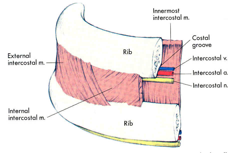
The intercostal muscles are two sets of muscles that are found between the ribs. Contraction of these muscles can either pull the rib cage up and out, or push the rib cage down and in. The muscles on the outside are called the external intercostal muscles and the ones on the inside are called internal intercostal muscles.
When you are breathing at rest the rib cage does not move at all. (I hope everyone reading this post is calm, relaxed and not hyperventilating in panic over upcoming exams….) As you are breathing at rest the only muscle involved is the diaphragm (see section above) as you are only moving about half a litre of air in and out with each breath. But there are situations in which this tidal volume has to increase and that is when the intercostal muscles come into their own.
The two sets of intercostal muscles are antagonistic – when one contracts the other relaxes.
If you need to take a big breath in, the external intercostals will contract at the same time as the diaphragm. The external intercostals pull the ribcage up and out, thus increasing even further the volume of the thorax, thus dropping the air pressure even more in the thorax, allowing more air to come in. When you come to breathe out, the external intercostal muscles will relax and gravity will allow the ribcage to fall back down to its original position.
But I hear you say…. “What happens if you are lying down or upside down? How can the ribcage get back to its original position without the help of gravity?” Well don’t worry – you have the internal intercostals which in extreme situations will contract during exhalation to push the ribcage down and in…
I suggest you draw up a table to summarise the process of breathing. Give inhalation and exhalation a column each, and the rows of the table should be diaphragm, external intercostals, internal intercostals… Tweet me a photo of your table if you want me to have a look…
