Feedback on Zondle Biology revision challenge part 2

The questions in this revision test were more challenging than last time.  I hope that players found them interesting and useful.

The plant transport questions at the start were well answered overall.  Osmosis is the only way water can ever cross a cell membrane and although active transport does occur in the root hair cells (pumping mineral ions such as nitrates into the cell against the concentration gradient), water cannot be directly pumped against its concentration gradient using energy from respiration.

The cloning questions were difficult but I think the low scores here were perhaps more to do with problems with my school wifi than with your abilities to answer them!  Micropropagation is the way that you learned when a small part of a plant is cut out, sterilised, washed and then added to a culture medium that triggers cell differentiation.  You probably did this experiment with explants from a cauliflower.  The aim was to produce whole new plants from these small explants.  This technique could not work with animals simply because animal’s bodies contain many more types of tissue and have a more complex internal architecture that requires a much more sophisticated genetic programme of development.

I want to talk about a few questions in the latter stages of the test that were not well answered.  I am sure there is plenty you can all learn from these.

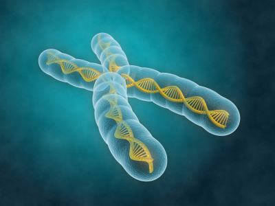
The first was the one that asked you what was meant by a “diploid cell”.  More than half of you thought that  diploid meant having 46 chromosomes.  This is almost a trick question because of course in humans, diploid cells will have 46 chromosomes.  But diploid can be applied to any cell that has chromosomes found in homologous pairs.  The number 23 is only important to humans as for our species it is the number of homologous pairs of chromosomes found in our diploid cells.  Different species have differing numbers of pairs of chromosomes, some less than the number in humans but in many species they have more.

The second big idea question was the true or false question on whether energy is recycled in the ecosystem like carbon atoms.  It is vital you understand that there is absolutely no recycling of energy ever in an ecosystem.  Energy enters in the form of light energy being trapped by plants in photosynthesis and all this energy ultimately ends up as heat energy in the atmosphere.  To find out the details of how it gets there, please read the relevant sections on my blog.  Try the tag energy from the Tag cloud on the right of the screen.

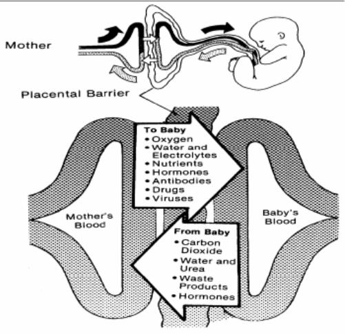
There was one question in the quiz which not a single player answered correctly and it is the one about which type of cells produce antibodies.  Antibodies are made from a cell called a plasma cell.  Plasma cells secrete antibodies in large numbers to combat an infection.  Plasma cells are descended from B lymphocytes that have been activated by the presence of antigen.  This clonal selection theory is one of the most complicated bits in iGCSE Biology so make sure you have looked carefully at it.  The final question was about active v passive immunity.  This is not specifically mentioned in the specification so perhaps is a bit mean to include but if you can understand it properly, you understand how immunity works.  Passive immunity is the name for when antibodies are transferred, perhaps across the placenta for a foetus or in an injection as an adult.  Antibodies are made of protein and so do not exist for long in the blood – after a month or two they will all have been broken down and cleared from the blood.  So passive immunity cannot give long-lasting protection.  Active immunity is when memory cells are produced via a clonal selection response.  These memory cells can survive for an entire lifetime and so do provide long lasting protection.

By far the biggest thing you can learn from this quiz however was about virus structure.  I asked you whether “viruses are made from a different kind of cell not found in animals or plants – true or false.”  Almost everyone went for false but remember this can’t be correct:  viruses are definitely not made of cells!  They are much simpler than even the simplest cell and just consist of a protein coat with some genetic material (DNA or RNA) inside.  No cell membrane, no cytoplasm, no metabolism – just two chemicals associated into one simple particle.

Anyway I hope you enjoyed the quiz – look out for the next one on my Twitter feed and please use the comment facility on this blog to get in touch if you have any questions or want more explanations.

Buttler, Humbug and Cricket’s Moral Code

I may be a single, dissenting voice in the “controversy” in the Joss Buttler run out yesterday, certainly from reading some of the utter tosh in the newspapers this morning.  Ashley Giles called the dismissal “unsavoury” along with a whole host of rentaquote ex-players (who all work in the media so have to add to the media storm) and I have just read the MailOnline’s report that says that “the run out is legal but it is not something anyone would want to see a youngster repeat”.  Well speaking as someone who has coached “youngsters” (what a strange word) for over 20 years and who views the moral code of cricket as one of the bedrocks of the game, I would have no problem with any of my cricketers doing exactly the same.

What I find unsavoury is batsmen backing up way out of the crease before the bowler even approaches his delivery stride.  I have a word for this – it’s called cheating.  When I am umpiring schoolboys I loudly tell the batsmen that he should know that I will always give him out with no hesitation if he is run out in this way.  You see it all the time as an umpire, almost every game and almost always from the better batsmen, those who have played the so called “higher levels” of schoolboy cricket.

For what it’s worth, I think the Sri Lankans deserve tremendous credit for giving him two warnings before running him out.  The laws were altered a few years ago to give batsmen more of an advantage (I won’t bore you with details) and so comparisons with the “Mankad” incident are totally erroneous and irrelevant.  The idea is that this idea of warning should be removed from the spirit of cricket and of people are cheating, run them out and they might not do it again.  Buttler is not guilty of dozily walking out of his crease in some kind of daze as some commentators are insinuating.  He is making a totally calculated decision that it is worth the risk to try to gain an unfair advantage, a risk not allowed in the laws of the game, in the hope that he is unlikely to ever get taken up on it.  Well now he has, and perhaps he will stop cheating when he is batting and other younger cricketers will follow……  Rant over.

Mutation – Grade 9 Understanding for IGCSE Biology 3.34 3.37B

Mutations are changes in the DNA content of a cell.  There are various ways the DNA of a cell could change and so mutations tend to be grouped into two main categories:  chromosomal mutations and gene mutations.

Image

Chromosomal mutation

This is a change in the number or length/arrangement of the chromosomes in the nucleus.  For example, people with Down’s syndrome have an extra copy of chromosome 21 giving them three chromosome 21s as opposed to the normal two.

(How many chromosomes in total will a person with Down’s syndrome have in each cell?)

Image

Chromosomal mutations are often found in tumour cells and so play a critical role in the development of various cancers.

Sometimes the number of chromosomes in a cell stays the same, but sections are deleted, duplicated or break off from one chromosome to attach elsewhere.  If this happens, this too would be classed as a chromosomal mutation.

Image

Image

Gene mutation

Gene mutations happen to change the sequence of base pairs that make up a single gene.  As you all know, the sequence of base pairs in a gene is a code that tells the cell the sequence of amino acids to be joined together to make a protein.  A gene then is the sequence of DNA that codes for a single protein.  If you alter the sequence of base pairs in the DNA by adding extra ones in, or deleting some or inverting them, this will alter the protein produced.

Image


A point mutation is a change to just one base within the gene – it occurs at a single point on the DNA molecule.

Mutations can happen at any time and occur randomly whenever the DNA is replicated.  But there are certain things that can increase the rate of mutation and so make harmful mutations more likely.  A mutagen is an agent that increases the chance of a mutation occurring.

a) Radiation can act as a mutagen

Some parts of the electromagnetic spectrum can cause mutations when they hit DNA molecules or chromosomes.  This is called ionising radiation and includes gamma rays, X rays and ultraviolet.  You probably know that the dentist goes out of the room whenever they take an X ray to protect themselves from repeated exposure to X rays and you all certainly know of the link between UV exposure and incidence of skin cancer.

b) There are chemical mutagens as well

Some chemicals can make the rate of mutation increase.  These are called chemical mutagens and a good example is the tar in tobacco smoke.  Tar can cause cancers to form wherever the cigarette smoke comes into contact with cells and this is because tar is a mutagen.  It makes mutations in the DNA much more likely and mutations are needed to turn a healthy cell into a cancer cell.


Role of the Amnion – Grade 9 Understanding for IGCSE Biology 3.12

Once the embryo has reached the uterus 7 days after fertilisation, it can implant into the thickened, sticky and blood-rich endometrium.  The implanted embryo grows into the uterine lining and starts to surround itself with a collection of membranes.  Some of these membranes develop into structures in the placenta, but one the amnion has a different function altogether.  The amnion produces a fluid called amniotic fluid that cushions the developing embryo and foetus right through pregnancy and to birth.

Image


The main advantage of having the developing embryo in a sac of amniotic fluid is that it protects the embryo by cushioning against blows to the abdomen.  It is also essential for allowing the foetus to move around inside the uterus thus allowing development of the muscular system.  The amniotic fluid enters the babies lungs and can promote normal development there.  The foetus will swallow amniotic fluid into its stomach and will produce urine into the amniotic fluid as well.  Disgusting I know, but that’s babies for you……..

Amniotic fluid contains stem cells.  In the future it may be possible to harvest these pluripotent cells and use them to create adult tissues for medical uses.

Role of the Placenta – Grade 9 Understanding for IGCSE Biology 3.11

There are two syllabus points in bold (only tested in paper 2) that refer to embryonic and foetal development.  The first asks you to understand the role of the placenta in supplying the developing foetus with nutrients and oxygen and the second concerns the role of amniotic fluid in protecting the developing embryo.

Placenta

The placenta is in many ways a remarkable organ.  It contains a mixture of maternal cells from the uterine lining and embryonic cells, but these cells from two genetically different individuals are capable of sticking together to form the placenta.  The placenta is only present in the uterus once an embryo has successfully implanted a week or so after fertilisation has happened in the Fallopian Tubes.  The placenta is linked to the foetus via the umbilical cord, a structure that contains an umbilical artery and vein carrying foetal blood to and from the placenta.

Image


There is a key idea here that is very important.  There is no mixing of maternal and foetal blood in the placenta.  This would be disastrous for both mother and baby for a whole variety of reasons.  The maternal blood is at a much higher pressure than the foetal blood and if the foetus were connected to the maternal circulatory system directly, its blood vessels would burst.  The foetus and mother can have different blood groups of course and you may now that some blood groups are incompatible and can trigger clotting.  So it is essential that there is never any mixing of blood.  But what happens in the placenta is that mother’s blood empties into spaces in the placenta and babies’ blood is carried by the umbilical artery into capillaries that are found in finger-like projections called villi.  This means there is a large surface area and a thin barrier between the two bloods and so exchange of materials by diffusion is possible.

The main function of the placenta then is to allow the exchange of materials between the foetal and maternal circulations.  The developing foetus inside its mother’s uterus has no direct access to oxygen nor food molecules of course yet both are needed to allow healthy development.  The foetus also needs a mechanism to get rid of the waste molecule, carbon dioxide that is being produced in all its cells all the time.  Until the kidneys mature fully the foetus also has to get rid of urea, another excretory molecule that could build up to toxic concentrations unless removed from the growing foetus.

Image

A few interesting points:

You will see that antibodies are small enough to cross the placenta.  This gives the baby a passive immunity that can protect it for a short time from any pathogens it encounters.

Drugs such as alcohol and nicotine can cross the placenta.  This is why it is so vital that pregnant mothers do not smoke and drink to ensure that the foetus’ development is not affected by these drugs.


Germination – Grade 9 Understanding for Biology GCSE 3.5 3.6

In the topic of sexual reproduction in plants, the final stage is often overlooked.  I think it is helpful for students to think of this topic in several distinct stages.

  • Flower Structure (hermaphrodite nature of most plants)
  • Pollination (self v cross pollination; wind v insect pollinated flowers)
  • Fertilisation (how does the pollen tube reach the egg cell to fertilise it?)
  • Seed and Fruit formation (what forms what after fertilisation)
  • Seed Dispersal (by animals, by wind, by water, by explosive means)
  • Germination

Once the seed has been dispersed there then follows a period of dormancy when nothing happens.  In latitudes such as the UK this often is there to delay germination until the following spring when growing conditions become more favourable.  The process of taking an inert seed and growing a new plant from it is called germination.

You don’t need to worry too much about the details of germination but there are a few vital parts of the process that GCSE candidates need to appreciate for A* marks.  Firstly you should know the structure of a typical seed.

Image

The seed coat or testa surrounds the seed and provides a tough waterproof container.  Inside there are the embryonic plant (composed of a plumule and radicle), one or two seed leaves called cotyledons and a storage tissue called endosperm.

Germination starts when the seed starts to take up water by osmosis.  There is an opening in the testa called the micropyle that allows water to move into the seed causing it to swell and thus rupture the seed coat to allow the embryo plant to emerge.

Image

Water entering the seed also activates the embryo plant such that it starts to release digestive enzymes such as amylase.  Amylase catalyses the digestion of starch into a simple sugar maltose.  The endosperm and cotyledons contain energy stores in the form of starch, lipids and proteins and as these get broken down by the various enzymes, they provide the energy for the early growth of the seedling.  The radicle emerges first and grows downwards (positive geotropism) and then the plumule or shoot grows upwards towards light (positive phototropism).  Remember that throughout the early stages of this growth the energy required comes from stored food molecules in the seed.  If you measure the mass of the plant during this phase, it would be decreasing.  Only when the first leaves emerge above ground and the plant can start the process of photosynthesis will the overall mass start to increase.

Image

Thoughts on Waiting for Godot at the Arcola ****

First comes a confession.  I have never seen this play before, never even been tempted to see this play before now and know nothing at all about Beckett other than his nationality and the century in which he wrote his plays.   As I have never written a theatrical review before now, this seems like too good an opportunity to miss…..   To be totally honest with you the reader, this is not really a “review” just a few jumbled thoughts early on a Sunday morning.

I suppose my “lack of previous” gives me the distinct advantage of having nothing with which to compare Simon Dormandy’s new production at the Arcola:  so I am not hung up on the age of the protagonists nor on the kind of headgear they wear.  I cannot compare the actors’ performances to Patrick Stewart or Ian McKellen but can give you a few thoughts on the play from someone who has seen little if any existentialist theatre.  The first thing to say is that I enjoyed the two and a half hours I spent in the actors’ company last night.  The set is dark, dusty and powerful.  I really liked the way the tree (and indeed one of the cast) seemed to grow out of the rubble.  The play is famous for its lack of narrative but I found the performances of all five members of the cast compelling in their different ways.  Tom Stourton’s Estragon manages to bring a weary resignation to his role and at times in the play he convinced me he was a man on the edge of desperation and total despair.  The highlights for me were when he could inject some physical comedy into his interactions with the rather nerdy Vladimir, played by Tom Palmer.  Pozzo steals the stage in the first act and then, after the interval, his return marks the start of the darkest and bleakest section of the play when the fabric of reality starts to crumble even more.

I am sure Beckett intended the play to provoke rather than answer questions, but I was left wondering what he was trying to say about the uncertainty of memory, the fluidity of time and the perhaps the ultimate futility of the struggle.  The relatively young age of the tramps does pose one major challenge for this production.  If Vladimir and Estragon are old men then it is easier to believe that their ultimately fruitless wait might be better ended by a quick hanging from the tree.  But I think we have a strongly ingrained preconception of the power of youth to change its circumstances.  Is it possible that two young men in their twenties are really living such a bleak and worthless existence as to lose all hope?  Do all their uncertainties about memory on display happen to younger people as well?

I know nothing about the religious background to this play but the similarity of the tree to a cross and the repeated references to the crucifixion in the text seem too much of a coincidence to me.  Is there a message hidden in here about the true nature of salvation?

There are groups going from school in the next few weeks to see this play and I would recommend it to anyone.  If you can provide any answers to the questions this production so successfully poses, perhaps you could let me know?  A quick tweet with what it is all about would help me.  It might save me my own long wait.

Understanding the Eye to Grade 9 at GCSE Biology (part 2) 2.91 2.92

In the first blog post on this series, I described the pupil reflex in the eye.  If you remember this involved the circular and radial muscles in the iris contracting and relaxing in an antagonistic fashion to alter the size of the pupil.  You should understand why the pupil size needs to altered and what state the two sets of muscles are in varying light intensities.

But there is a second reflex in the eye totally separate from the pupil reflex and it is to do with focusing.  This reflex is sometimes called accommodation but as this is a word I can’t spell, I prefer to call it focusing…. The retina at the back of the eye contains the photoreceptors.  There are two types of photoreceptor in the retina (rods and cones) and these are individual cells that can detect the light and then send a nerve impulse in the optic nerve that goes to the brain.

Focusing in the Eye (this is quite complicated and needs careful, slow reading)

When the eye views objects from differing distances away, the degree the light has to be bent to produce a focused image will vary.  Light coming from near objects will be diverging (the rays will be moving away from each other) and so to focus the light onto the retina, a large amount of bending (or better still refraction) will be needed.  Light rays coming from far away objects are almost parallel when they hit the eye so the degree of refraction required is much less.

Eye-focus-final-3000X2000

How can varying degrees of refraction be achieved in the eye?

Well as the diagram above shows, this is brought about by changing the shape of the lens.  A short fat lens will refract (or bend) the light more than a long thin one.  (If you want an explanation for why this is, you need to ask a Physics teacher – it is to do with the angle of curvature of the lens and the refractive indices of the liquids in the eye compared to the lens…….)

The lens in its default state is short and fat.  This means that with no tension pulling it out of shape it will adopt the short fat shape suitable for viewing near objects.

How can the lens be pulled out of its default short fat shape?

Now this is the bit where people get confused.  Read this section really carefully, check with your own notes and revision notes and make sure you have got this all the right way round!  Here goes…..

There is a ring of muscle that surrounds the lens in the eye called the ciliary muscle.  (Please make sure you don’t confuse this with the circular muscles in the iris)  The ciliary muscle doesn’t attach to the lens directly but is attached to the lens via some strong and inelastic ligaments called the suspensory ligaments.   Tension in the suspensory ligaments can pull the lens from its default short, fat shape into the long this shape needed to view far away objects.

When the ciliary muscle contracts, it shortens.  This effectively moves it closer to the lens and so any tension in the suspensory ligaments is released as the ligaments go slack.  Slack ligaments mean the lens adopts its short fat shape.

When the ciliary muscle relaxes, this changes its position to increase the tension in the suspensory ligaments.  Taut suspensory ligaments (caused by the relaxed muscle) will pull the lens into a long thin shape.

You can easily see why people get confused here:  a contracted ciliary muscle leads to slack suspensory ligaments and vice versa.

Image

One way you can improve your understanding is to be really precise with your use of language.  The ciliary muscle is a muscle (no honestly it is) and as you know, muscles can either contract or relax.  Suspensory ligaments cannot contract or relax but their tension can be altered from taut (loads of tension) to slack.

So to summarise this complex sequence of events:

Looking at a far object

  • Lens needs to be long and thin
  • as light rays are almost parallel as they hit the eye
  • and so require little bending.
  • To pull the lens long and thin requires
  • suspensory ligaments to be taut
  • and this is achieved by the ciliary muscle relaxing.

Looking at a near object

  • Lens needs to be short and fat
  • as light rays are diverging as they hit the eye
  • and so require a lot of bending.
  • The lens will adopt a short, fat shape with
  • no tension in the suspensory ligaments (the ligaments are slack)
  • and this is achieved by contracting the ciliary muscle.

You can easily check your understanding here because it is much more tiring on the eye to look at a near object.  If you sit on a sunny beach after all your GCSEs are finished, staring out to sea in a contemplative manner wondering how you managed to work so hard through the revision period, you could continue like this for hours.  But if you try staring at your finger a few centimetres from your face for even a few seconds, your eye starts to tire.  In the former scenario the ciliary muscle is relaxed and so not expending any energy but in the latter, the ciliary muscle is contracted, using energy from respiration and so can get tired.

Please comment me on this blog post with any questions – I will do my best to respond to anyone who gets in touch.

Good luck and keep working hard!

Understanding the functioning of the Eye to Grade 9 for Biology IGCSE (part 1) 2.91, 2.92

There are two reflex responses in the eye that you need to fully understand for A* levels at iGCSE.   It is really easy to get them confused but I am going to put on consecutive blog posts so you can see the similarities and differences easily.

The first is a reflex called the “Pupil Reflex” which is to ensure an appropriate amount of light enters the eye in both bright and dim light.  The only structure in the eye involved in the Pupil Reflex is the Iris.  The second reflex explained in part 2 is the “Focusing Reflex” (or sometimes Accommodation) which makes sure that light entering the eye from objects at different distances away is focused correctly onto the retina.  The structures involving in Focusing are the Lens, Ciliary Muscle and Suspensory Ligaments.

The Pupil Reflex

1) Why do we need a pupil reflex?

The eye has evolved a mechanism to ensure that the amount of light entering the eye can be adjusted.  In bright light you need to limit the amount of light to prevent the light damaging the light-sensitive cells in the retina (a process called “bleaching”) and this is done by making the pupil at the front of the eye small.  A small pupil would be useless for vision in low light intensities as then not enough light would get to the retina and vision would be very poor.  So in dim light (low light intensities) the pupil is enlarged to allow a maximal amount of light into the eye.

2) What is the Pupil?

The pupil isn’t really a structure at all as it is simply a circular hole in the iris.  The iris is a coloured muscular disc at the front of the eye.

Image

The iris has two sets of antagonistic muscles in it that can contract or relax to change the diameter of the pupil.  There are radial muscles arranged like the spokes of a bicycle tyre and also circular muscles in the iris as shown in the diagram below.

S182_5_005i


3) How do the muscles in the iris bring about the pupil reflex?

Remember muscles can only contract or relax.  When the radial muscles contract (shorten) they will pull the iris into a narrower shape so the pupil gets much wider.  When the circular muscles contract, they will squeeze the pupil smaller so the pupil will narrow.

pupil_reflex_med


So you need to basic understand the state of these two sets of antagonistic muscles in both bright and dim light.

Bright light – circular muscles contracted, radial muscles relaxed, pupil small

Dim light – circular muscles relaxed, radial muscles contracted, pupil large

Commentary on Zondle GCSE Biology Revision challenge 1 questions

I hope that those of you who played the GCSE Biology revision challenge this afternoon enjoyed the process.  I would welcome comments on this blog post along the lines of www (what went well) and ebi (even better if)….

The questions were grouped into several topic areas.

Questions 1 to 4 were on thermoregulation.  Understanding vasoconstriction, vasodilation and sweating are the key things here and if you haven’e done already, I would read my blog post on this topic.

https://pmgbiology.wordpress.com/2014/05/29/skin-a-understanding-for-igcse-biology/

Questions 5 – 9 were on plant transport and these were well answered by almost all players.  Remember that phloem sieve tubes move sucrose and amino acids around the plant.  Water and minerals are transported in xylem vessels of course, but the other distractor answers included various polymers (starch and proteins) that are made in photosynthesis in the leaves but which are too large and insoluble molecules to be transported in phloem.

Questions 10-15 were all on the bacteria in the Nitrogen cycle.  This is a tricky topic but one that rewards patient work by candidates to master it. In reality the Nitrogen cycle is not difficult to understand but it is easy to muddle the names and roles of the four types of bacteria involved.  Again there are a couple of blog posts on Nitrogen cycle that I would encourage you to read….

https://pmgbiology.wordpress.com/2014/04/12/bacteria-of-the-nitrogen-cycle-a-understanding-for-gcse-biology/

https://pmgbiology.wordpress.com/2014/04/12/nitrogen-cycle-for-igcse-biology/

Questions 16-23 were on digestive systems.  These were generally well answered although many players didn’t appreciate that peristalsis doesn’t just happen in the oesophagus: it is the process that moves the food along the entire length of the gut tube from top of oesophagus to the end of the rectum.  The role of the lacteal in transporting fatty acids and glycerol away from the villi in the small intestine is also one of the trickier topics here.  Amino acids and sugars diffuse into the blood capillaries in the villus but fatty acids and glycerol (the products of digestion of lipids) don’t go into the capillaries but instead into a separate vessel called a lacteal. This forms part of the lymphatic system and the liquid formed ends up back in the blood but effectively bypasses the liver, preventing the cells in the liver being overloaded with fatty acids following a fatty meal.

Questions 24-29 were on the heart and circulation.  There were quite a few incorrect answers here but perhaps this was because enthusiasm levels were dropping….  The flow of blood through the heart is an important topic to appreciate – into RA through vena cava, then into RV through right AV valve, then into PA through semilunar valve, then to lungs, back from lungs in pulmonary veins, into LA, through Left AV valve into LV, then into aorta through the aortic semilunar valve…..

The heart strings in the heart (chordae tendinae) are commonly misunderstood.  They play no role at all in opening or closing the AV valves (this is done simply by the balance of blood pressures in atrium and ventricle) but do provide tension to stop the valve “blowing back” and thus opening when the ventricle contracts.  Have a look at pictures of a real heart dissection to see that these tendons attach to the valve flaps and ensure they cannot blow open when the pressure in the ventricles rise during ventricular contraction.  Ask me for more detail if this doesn’t make sense.