
Tagged: 2.88
Understanding the Eye to Grade 9 at GCSE Biology (part 2) 2.91 2.92
In the first blog post on this series, I described the pupil reflex in the eye. If you remember this involved the circular and radial muscles in the iris contracting and relaxing in an antagonistic fashion to alter the size of the pupil. You should understand why the pupil size needs to altered and what state the two sets of muscles are in varying light intensities.
But there is a second reflex in the eye totally separate from the pupil reflex and it is to do with focusing. This reflex is sometimes called accommodation but as this is a word I can’t spell, I prefer to call it focusing…. The retina at the back of the eye contains the photoreceptors. There are two types of photoreceptor in the retina (rods and cones) and these are individual cells that can detect the light and then send a nerve impulse in the optic nerve that goes to the brain.
Focusing in the Eye (this is quite complicated and needs careful, slow reading)
When the eye views objects from differing distances away, the degree the light has to be bent to produce a focused image will vary. Light coming from near objects will be diverging (the rays will be moving away from each other) and so to focus the light onto the retina, a large amount of bending (or better still refraction) will be needed. Light rays coming from far away objects are almost parallel when they hit the eye so the degree of refraction required is much less.

How can varying degrees of refraction be achieved in the eye?
Well as the diagram above shows, this is brought about by changing the shape of the lens. A short fat lens will refract (or bend) the light more than a long thin one. (If you want an explanation for why this is, you need to ask a Physics teacher – it is to do with the angle of curvature of the lens and the refractive indices of the liquids in the eye compared to the lens…….)
The lens in its default state is short and fat. This means that with no tension pulling it out of shape it will adopt the short fat shape suitable for viewing near objects.
How can the lens be pulled out of its default short fat shape?
Now this is the bit where people get confused. Read this section really carefully, check with your own notes and revision notes and make sure you have got this all the right way round! Here goes…..
There is a ring of muscle that surrounds the lens in the eye called the ciliary muscle. (Please make sure you don’t confuse this with the circular muscles in the iris) The ciliary muscle doesn’t attach to the lens directly but is attached to the lens via some strong and inelastic ligaments called the suspensory ligaments. Tension in the suspensory ligaments can pull the lens from its default short, fat shape into the long this shape needed to view far away objects.
When the ciliary muscle contracts, it shortens. This effectively moves it closer to the lens and so any tension in the suspensory ligaments is released as the ligaments go slack. Slack ligaments mean the lens adopts its short fat shape.
When the ciliary muscle relaxes, this changes its position to increase the tension in the suspensory ligaments. Taut suspensory ligaments (caused by the relaxed muscle) will pull the lens into a long thin shape.
You can easily see why people get confused here: a contracted ciliary muscle leads to slack suspensory ligaments and vice versa.

One way you can improve your understanding is to be really precise with your use of language. The ciliary muscle is a muscle (no honestly it is) and as you know, muscles can either contract or relax. Suspensory ligaments cannot contract or relax but their tension can be altered from taut (loads of tension) to slack.
So to summarise this complex sequence of events:
Looking at a far object
- Lens needs to be long and thin
- as light rays are almost parallel as they hit the eye
- and so require little bending.
- To pull the lens long and thin requires
- suspensory ligaments to be taut
- and this is achieved by the ciliary muscle relaxing.
Looking at a near object
- Lens needs to be short and fat
- as light rays are diverging as they hit the eye
- and so require a lot of bending.
- The lens will adopt a short, fat shape with
- no tension in the suspensory ligaments (the ligaments are slack)
- and this is achieved by contracting the ciliary muscle.
You can easily check your understanding here because it is much more tiring on the eye to look at a near object. If you sit on a sunny beach after all your GCSEs are finished, staring out to sea in a contemplative manner wondering how you managed to work so hard through the revision period, you could continue like this for hours. But if you try staring at your finger a few centimetres from your face for even a few seconds, your eye starts to tire. In the former scenario the ciliary muscle is relaxed and so not expending any energy but in the latter, the ciliary muscle is contracted, using energy from respiration and so can get tired.
Please comment me on this blog post with any questions – I will do my best to respond to anyone who gets in touch.
Good luck and keep working hard!
Understanding the functioning of the Eye to Grade 9 for Biology IGCSE (part 1) 2.91, 2.92
There are two reflex responses in the eye that you need to fully understand for A* levels at iGCSE. It is really easy to get them confused but I am going to put on consecutive blog posts so you can see the similarities and differences easily.
The first is a reflex called the “Pupil Reflex” which is to ensure an appropriate amount of light enters the eye in both bright and dim light. The only structure in the eye involved in the Pupil Reflex is the Iris. The second reflex explained in part 2 is the “Focusing Reflex” (or sometimes Accommodation) which makes sure that light entering the eye from objects at different distances away is focused correctly onto the retina. The structures involving in Focusing are the Lens, Ciliary Muscle and Suspensory Ligaments.
The Pupil Reflex
1) Why do we need a pupil reflex?
The eye has evolved a mechanism to ensure that the amount of light entering the eye can be adjusted. In bright light you need to limit the amount of light to prevent the light damaging the light-sensitive cells in the retina (a process called “bleaching”) and this is done by making the pupil at the front of the eye small. A small pupil would be useless for vision in low light intensities as then not enough light would get to the retina and vision would be very poor. So in dim light (low light intensities) the pupil is enlarged to allow a maximal amount of light into the eye.
2) What is the Pupil?
The pupil isn’t really a structure at all as it is simply a circular hole in the iris. The iris is a coloured muscular disc at the front of the eye.
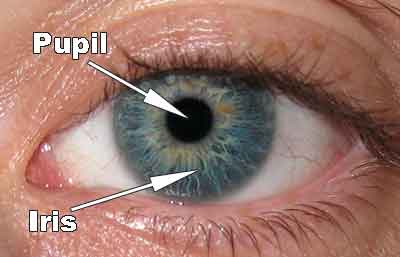
The iris has two sets of antagonistic muscles in it that can contract or relax to change the diameter of the pupil. There are radial muscles arranged like the spokes of a bicycle tyre and also circular muscles in the iris as shown in the diagram below.

3) How do the muscles in the iris bring about the pupil reflex?
Remember muscles can only contract or relax. When the radial muscles contract (shorten) they will pull the iris into a narrower shape so the pupil gets much wider. When the circular muscles contract, they will squeeze the pupil smaller so the pupil will narrow.

So you need to basic understand the state of these two sets of antagonistic muscles in both bright and dim light.
Bright light – circular muscles contracted, radial muscles relaxed, pupil small
Dim light – circular muscles relaxed, radial muscles contracted, pupil large
