Category: Section 2: Structures and Functions in Living Organisms
Bile: A* Grade 9 Understanding for IGCSE Biology 2.30 2.31
The liver is the largest internal organ and plays over 500 different roles in the body. Many functions are to do with the processing of various chemicals such as carbohydrates, amino acids and lipids. The liver also removes alcohol and other drugs from the bloodstream: this is why alcoholics often suffer from liver disease.
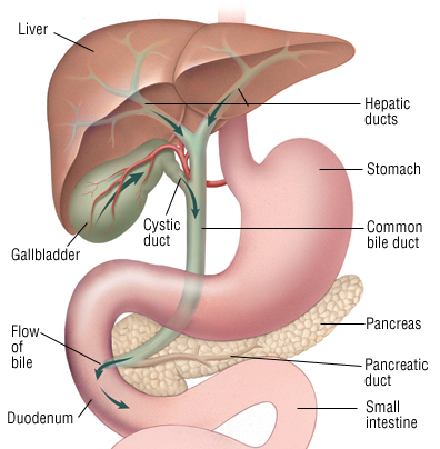
But one function of the liver that you need to understand in detail concerns its role in the digestive system. The liver cells produce a green liquid called bile which is stored in a sac underneath the liver called the gall bladder. Bile can pass from the gall bladder down the bile duct and as shown in the diagram below, it then mixes with the contents of the duodenum (small intestine) soon after the acidic chyme leaves the stomach.
What is in bile?
Bile contains a mixture of chemicals. It has an alkaline substance (hydrogencarbonate ions) which helps to neutralise the chyme as it leaves the stomach. Remember the pH of the stomach contents is around pH1-2 and in the duodenum, there is a pH of around pH7.5. The difference in pH is due to the alkali present in bile and pancreatic juice. Bile also contains excretory molecules called bile pigments. These are waste molecules from the liver that have been made from the breakdown of haemoglobin. And finally there are the bile salts. These play an important role in the digestion of lipids in the duodenum.
How do bile salts improve digestion of lipids in the duodenum?
The duodenum is where many digestive reactions happen in the body. This is because the pancreatic juice contains many enzymes, all of which catalyse a specific digestive reaction. One such enzyme is lipase and this enzyme catalyses the following reaction:
lipids + water ——> glycerol and fatty acids
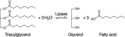
Fatty acids and glycerol are small enough molecules to be absorbed into the villi in the ileum. They pass into the lacteal in the centre of each villus to be carried around the body in the lymphatic system.
But the problem is that in the duodenum, the lipid molecules will exist as large droplets. Large droplets of fat/oil will have a reduced surface area for lipase to bind to and so the rate of digestion of the lipid would be slow. But bile salts interact with the lipid droplet causing a few large droplets to be broken down into dozens of tiny droplets. This is called emulsification and while it does not chemically alter the lipid, it does make it easier for lipase to break it down. Lipase and bile salts together break down lipids much faster than lipase alone.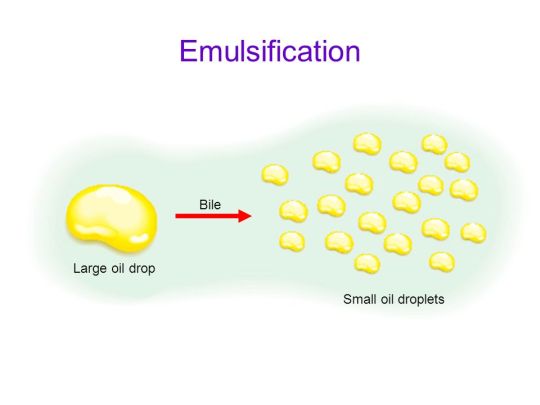
Final key point: there are no digestive enzymes in bile. But in spite of this, bile plays a crucial role in the digestion of lipid droplets in the duodenum.

Small Intestine: Grade 9 Understanding for IGCSE Biology 2.32
The first part of the small intestine, called the duodenum is principally involved in digestion. Large insoluble food molecules such as proteins, lipids and starch are chemically broken down into smaller molecules in reactions catalysed by digestive enzymes.
This post will look at the longer regions of the small intestine, the jejunum and ileum. There are some digestive reactions that happen here but the main function of these parts of the intestine is the absorption of the smaller products of digestion into the body.

You should understand already which molecules are produced as products of digestion: glucose from the digestion of carbohydrates, amino acids from the breakdown of proteins and fatty acids and glycerol from the digestion of triglyceride lipids. (see my post on digestion) These then are the molecules that diffuse from the intestine into the body in the small intestine.
How is the structure of the small intestine adapted for absorption?
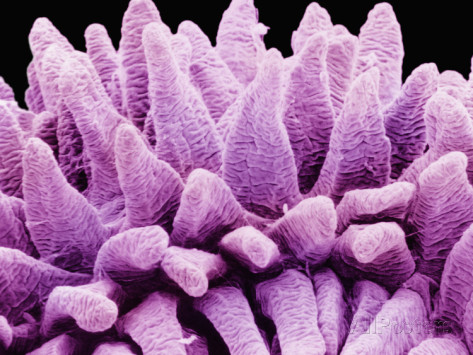
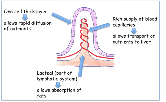
The main idea here is that the lining of these parts of the small intestine has a very large surface area. The intestine is long, the wall is ridged and the lining (called the epithelium) has many thousands of tiny projections called villi. Each villus is 1-3mm long but the effect is to increase the surface area for absorption by many hundreds of times.
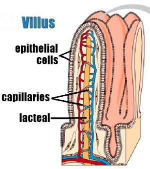
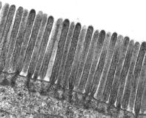
These epithelial cells have a cell membrane which is folded into many thousands of tiny structures called microvilli. Microvilli (or a brush border) can only be seem with an electron microscope and act to increase the surface area still further.
The cells that line the villus are called epithelial cells and are found in a layer that is just one cell thick. This reduces the distance the products of digestion have to move across to be absorbed.
Each villus contains a dense network of blood capillaries. This means that glucose and amino acids can easily diffuse into the blood and then be taken away from the small intestine to the liver in the hepatic portal vein. There is also a blind-ended single tube called a lacteal in each villus. This tube forms part of the lymphatic system and is used to transport fatty acids and glycerol away from the small intestine.
Stages in Processing Food: A* understanding of animal nutrition for IGCSE (no longer in specification)
Most animals including humans feed by a process called holozoic nutrition. This means that the animal has a gut tube (alimentary canal) that runs through its body and the animal has a mouth at one end (this is where the food goes in), an anus at the other end where undigested food (faeces) passes out.

Ingestion: the first stage of feeding involves food being taken up into the mouth (not too complicated to understand I hope…)
Most food is made up of very large molecules (macromolecules) such as starch, proteins and lipids. These molecules are too large to be absorbed from the gut tube into the blood. So the second stage of feeding involves chemically breaking down these large food molecules into smaller solubles molecules that can be absorbed.
Digestion: the chemical breakdown of large insoluble food molecules (e.g. proteins/starch/lipids) into smaller soluble molecules (amino acids/sugars/fatty acids and glycerol) that can be absorbed. Digestion is a chemical process and every digestive reaction is catalysed by a specific enzyme. Almost all these digestive enzymes are secreted into the gut tube and mixed in with the food. Please see a later post on Digestion to get a full understanding of these reactions.
Absorption: the small products of digestion are then absorbed into the blood stream. This process occurs almost exclusively in a region of the small intestine called the ileum. The structure of the ileum is beautifully adapted for efficient absorption – see a later post on Absorption for full details!
Assimilation: this is the stage of processing food that is sometimes left out. This is because it does not happen in the alimentary canal but instead in the body cells of the animal. The small soluble products of digestion (glucose, amino acids, fatty acids etc.) are taken up into cells and used to build the animal’s own macromolecules – proteins/lipids/glycogen etc. Assimilation is a term for how small molecules are built up and used in the cells of the animal.
Egestion: this is the final stage and involves undigested food, mostly cellulose in humans, being passed out of the anus as faeces at the end of the alimentary canal.
Don’t confuse egestion with excretion….. Egestion is simply passing out undigested food from the end of the large intestine. If you think of your body like a tube of polos (you know I do…) then the stuff that comes out of the large intestine has never actually been inside your cells. It is mostly cellulose and other plant fibres that went into the mouth a day or two ago, haven’t been digested nor absorbed and so come out the other end. Excretion on the other hand is the process of removing waste molecules that have been made inside cells. So the lungs removing carbon dioxide, the kidneys taking urea out of the blood to make urine – these are examples of excretion.
A slightly lavatorial way to end the post but important nonetheless: if you are talking about urine production, this is excretion, but “number twos” or “Richards” (look up cockney-rhyming slang for EAL speakers) are not excretion, they are egestion….
The Human Alimentary canal: Grade 9 Understanding for IGCSE Biology 2.27
A human body is in many ways rather like a packet of polo mints. These are famous mints in the UK for having a hole in the middle. Our body is divided into segments (rather like the packet of polos) and we have a tube that runs through the middle of us. This tube is called the Alimentary Canal (or Gut) and it’s function in the body is the digestion and absorption of food molecules (see later post on “Stages of Processing Food”)

The Alimentary Canal is divided into specialised regions, each with its own particular range of functions to do with the processing of food. You are required to understand a little about some of these organs and their functions.

The first thing is to make sure you can label a diagram of the human digestive system such as the one shown above. Check that you could accurately identify the following structures:
mouth, tongue, teeth, salivary glands, oesophagus, stomach, liver, gall bladder, bile duct, pancreas, pancreatic duct, duodenum, ileum, colon, appendix, rectum, anus
Here is a good diagram to use to check your labelling of the human digestive system

Functions
1 Mouth
The mouth is actually the name for the opening at the top of the alimentary canal rather than the chamber behind. If you want to be really precise, you should call this chamber containing the tongue and teeth by its proper name, the buccal cavity. The mouth is the opening that allows an animal to ingest food. In the buccal cavity, the teeth can chop up the food into smaller pieces and the tongue can move the food into a ball (bolus) for swallowing. The food is tasted in the buccal cavity and there are many chemoreceptors on the tongue and in the nasal cavity that perform this function. There are three sets of salivary glands around the buccal cavity and these secrete a watery liquid, saliva to mix with the ingested food. Saliva is alkaline to help protect the tooth enamel from acidic decay by bacteria but also contains a digestive enzyme, salivary amylase that begins the process of digestion of starch in the mouth. Salivary amylase catalyses the hydrolysis reaction in which starch, a polysaccharide is digested into the disaccharide maltose.
2 Oesophagus
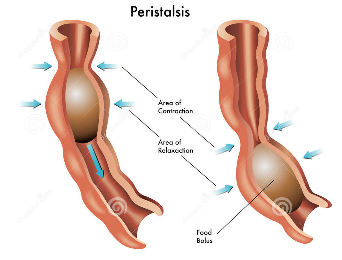
The oesophagus is the tube that carries the ball of food from the back of the throat through the thorax and down into the stomach. The Alimentary canal has layers of muscle in its wall throughout its entire length. These layers of smooth muscle can contract and relax in an antagonistic fashion to push the bolus along the tube. There are two main types of smooth muscle in the wall of the Alimentary canal – circular fibres are arranged around the circumference of the tube and longitudinal fibres are arranged along the length of the tube. These waves of alternate contraction and relaxation are called peristalsis.

3 Stomach
The stomach is a muscular storage organ that keeps food in it for around 3-4 hours before squirting it out in small amounts into the duodenum. The muscle layers in the stomach wall churn the food and mix it with the secretions from the stomach lining. These secretions are called gastric juice and contain a mixture of hydrochloric acid, mucus and a digestive enzyme pepsin. The acid makes the gastric juice overall very acidic, around pH 1.5. This acidity forms part of the non-specific defences of the body against bacteria as the extreme pH kills almost all bacteria in the food. The mucus is important as it protects the cells lining the stomach from the acidity. Pepsin is a digestive enzyme that starts the digestion of protein. It catalyses a hydrolysis reaction in which proteins are broken down into smaller molecules called polypeptides. Pepsin is an unusual enzyme in that it has an optimum pH of around 1.5.
4 Small Intestine
I will write a whole post on the small intestine later this week as there is plenty for you to understand about this part of the alimentary canal. All I will say here is that it is divided into the duodenum which is where almost all the digestion reactions take place and the ileum which is adapted for efficient absorption of the products of digestion into the blood. (see post later in the week if you want to find out more….)
5 Large Intestine
The majority of the large intestine is made up of an organ called the colon. The colon has a variety of functions. It is where water from all the various secretions is reabsorbed back into the blood, thus producing a solid waste called faeces. (Water that you drink tends to be absorbed through the stomach lining much earlier in the alimentary canal) There are also a few mineral salts and vitamins absorbed into the bloodstream in the colon. The colon is also home to a varied population of bacteria, the so called gut flora. Faeces is stored in the final part of the large intestine that is called the rectum.
6 Pancreas
The pancreas is not part of the alimentary canal (although I am not sure the person who wrote the specification appreciated that….) It is an example of what is called an accessory organ for the digestive system. The pancreas is a really interesting organ as it contains different cell types that carry out two completely separate functions. The majority of the cells in the pancreas secrete a whole load of digestive enzymes into an alkaline secretion called pancreatic juice. There is a tube called the pancreatic duct that carries the pancreatic juice and empties it into the duodenum where it can mix with the acidic chyme coming out of the stomach.
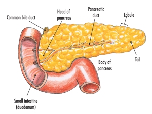
There are small clusters of a different kind of cell found in the pancreas. These are the islets of Langerhans that secrete the hormones insulin and glucagon into the bloodstream. These two pancreatic hormones together regulate the blood glucose concentration.

Alveoli and Gas Exchange: Grade 9 Understanding for IGCSE Biology 2.46 2.48
The primary function of the lungs is to allow gas exchange to occur. Oxygen gas can diffuse into the blood from the air in the lungs. Oxygen of course is needed for the process of aerobic respiration that is happening in every cell all the time. Aerobic respiration produces carbon dioxide as a waste product. Carbon dioxide diffuses out the blood in the lungs into the air in the lungs. Hence the name gas exchange – one gas (oxygen) diffuses in, another (carbon dioxide) diffuses out.
This diagram above shows the bronchial tree – the branching network of tubes that carry air into the lungs. The trachea at the top branches into the right and left bronchi, then each in turn branch into smaller bronchi and finally into the smallest tubes called bronchioles. Bronchioles carry air into a cluster of tiny airsacs called alveoli (not ravioli as AZB told his F division today…)
Diffusion is the passive movement of molecules of a liquid or gas from a high concentration to a low concentration. So the first question is what ensures that there is an appropriate concentration gradient for each gas to diffuse?

In order to understand this, you have to remember that the blood going to the lungs is deoxygenated. The right ventricle pumps deoxygenated blood to the lungs in the pulmonary arteries. The tiny alveoli are then covered with capillaries and these join together to form the pulmonary veins. The pulmonary veins carry the oxygenated blood back to the left atrium of the heart. So the blood coming to the lungs will have a low oxygen concentration but a high carbon dioxide concentration.
How are the structure of alveoli adapted for efficient gas exchange?
- The alveoli in total provide a large surface area for the diffusion of oxygen and carbon dioxide. The total surface area of the alveoli in humans is approximately 90 m2 – the equivalent of two tennis courts…..
- The walls of the alveoli are very thin. The alveolus is lined with a single layer of cells, and of course the capillaries are also only one cell thick. So the distance for the diffusion of oxygen and carbon dioxide is very small (hence the rate of diffusion is very fast)
- The alveoli have a rich blood supply. Alveoli are lined by many capillaries.
- The surface of the alveolus is moist. Gas exchange surfaces are always moist as oxygen and carbon dioxide will diffuse more rapidly if they are dissolved in water.
- Alveoli also contain a cell that secretes surfactant. This molecule reduces the surface tension in the film of water that lines the alveolus, allowing air to move in and out more smoothly.

Respiration: Grade 9 Understanding for IGCSE Biology 2.34 2.36 2.37 2.38
I can’t believe that it is over year since I started posting about iGCSE Biology misconceptions and yet I have never written about Respiration. If there is one topic that students misunderstand more than any other (apart perhaps from genetics), this must be it…. So I am going to try to explain in a straightforward way what respiration is and why it is so important for life.
Life requires energy. Living cells are constantly doing things that use up energy: pumping molecules across their cell membranes, moving organelles around the cell, cell division, nerve cells sending electrical impulses around the body, muscle fibres contracting etc. etc. In every case, this energy comes from a metabolic process called Respiration. It is a series of chemical reactions, catalysed by enzymes and in some way, it happens in all cells.
So let’s start with a good definition. [Examiners are simple souls and often start questions with the classic “What is Respiration?”]
Respiration is a series of chemical reactions that happens inside cells in which food molecules (for example glucose) are oxidised to release energy for the cell.
My definition has to be a little vague because although glucose is found in all the equations for respiration, other food molecules can certainly be respired. And oxygen is only used in aerobic respiration. Many organisms can only respire without oxygen (anaerobic respiration) and some, such as humans can switch between aerobic and anaerobic depending on the conditions.

Aerobic Respiration happens for the most part in tiny organelles in the cytoplasm called Mitochondria. The diagram above shows the structure of a mitochondrion (I wouldn’t worry about learning it but perhaps you should be able to recognise the characteristically folded inner membrane?)
What are the differences between aerobic and anaerobic respiration in humans?
Well we have mentioned two already and there are others…..:
- Aerobic respiration requires oxygen, anaerobic does not.
- Aerobic respiration takes place in mitochondria, anaerobic only occurs in the cytoplasm.
- Aerobic respiration produces much more energy per glucose molecule than anaerobic – it is a more complete oxidation of the glucose, so much more energy is released.
- Anaerobic respiration produces lactic acid as a waste product (in humans) whereas in aerobic, carbon dioxide and water are the products
The summary equations for the processes are different as well.
Aerobic respiration:
word equation Glucose + Oxygen ——> Carbon Dioxide + Water
balanced chemical equation C6H12O6 + 6O2 ——> 6CO2 + 6H20
Anaerobic respiration in humans:
Glucose —–> Lactic Acid
Anaerobic respiration in Yeast (a single celled fungus):
Glucose —–> Ethanol and Carbon Dioxide
A couple of final points to note:
Anaerobic respiration in muscle cells does not produce carbon dioxide as a waste product (see the equation above…) Lactic acid is the only waste product. But lactic acid will accumulate in muscles and stop the muscle functioning properly so after a period of intense activity, lactic acid needs to be removed. How does this happen?
Lactic acid moves from the muscle in the blood and is transported to the liver. In the liver, the lactic acid is metabolised in an aerobic pathway that uses oxygen. This is why sprinters will always be breathing fast after the race, even when they are standing still. Their body needs extra oxygen to oxidise the lactic acid they have produced during the race. This extra oxygen is termed an oxygen debt and is the oxygen needed in the liver to fully oxidise lactic acid to carbon dioxide and water.
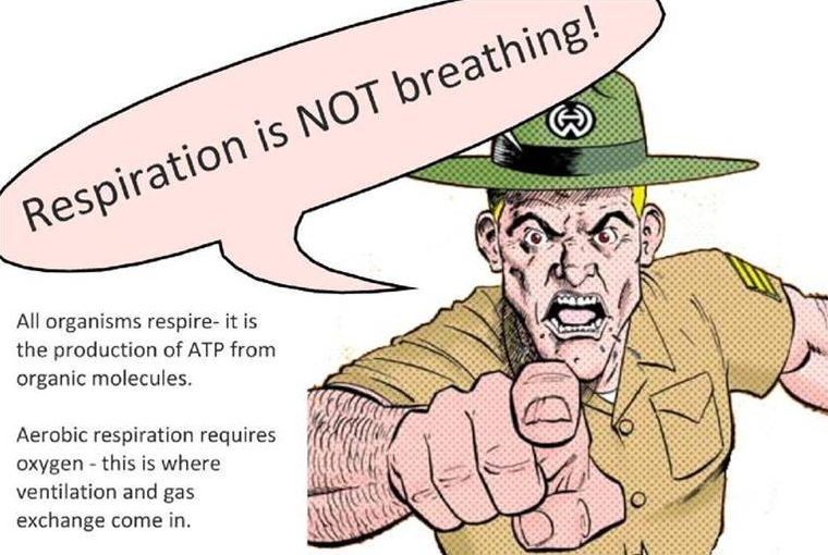
Finally, respiration is not the same as breathing. Our American cousins sometimes muddle these processes up but in this one case, the British way is much better…. Use the term ventilation for breathing – moving air in and out of the lungs – and reserve respiration for the chemical reactions that happen inside the cells to release energy.
Please leave a comment below if you find this post helpful or ask me about anything that isn’t clear….

Human Diet: Grade 9 Understanding for IGCSE Biology 2.24
All animals are heterotrophic. This means that they cannot make their own food molecules but need to get them from some external source. Humans get a variety of different food molecules from what they eat. Diet is a term for what an animal eats (and in a biological context has no associations with any attempt to lose weight or change body shape). A balanced diet is a combination of foods that provides the correct proportions of all the various food molecules for any particular individual at any particular stage of their life.
Components of a Balanced Diet
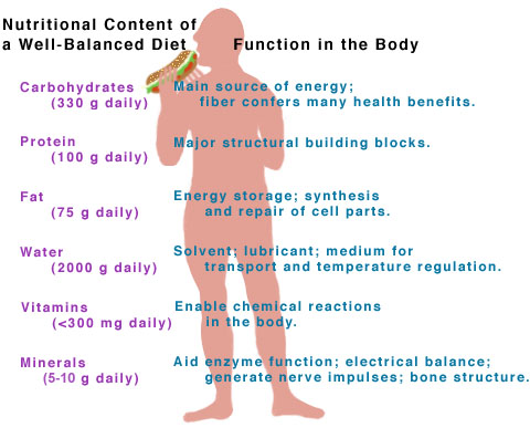
Carbohydrates
Carbohydrates are a family of molecules that includes sugars, starch and other polysaccharides. They contain C,H and O atoms only and their main function in the diet is to provide molecules that can be respired to release energy for cells. Carbohydrates are thus one of the main respiratory substrates in our diet. All sweet foods will contain sugars of course and starch-rich foods are vegetables like potatoes, pasta and rice. Starch is a polymer of glucose and so needs to be digested to glucose because it is too large a molecule to be absorbed in the small intestine.
Protein
Proteins are a family of macromolecules needed to build new cells and thus for growth. Like starch, Proteins are also polymers and thus get digested into their constituent monomers, in this case amino acids in the digestive system. Protein-rich foods include all meat and some pulses and beans. Proteins in the diet are needed to build muscle tissue, to form some components of cell membranes and to make all the enzymes that catalyse all the metabolic reactions in cells.
Lipids
Lipid is a general term for all fats and oils. Despite the popular misconception that fat is “bad” in our diet, in fact lipids are essential molecules in the diet. We need lipids as a respiratory substrate, for long term energy storage in adipose tissue under the skin and for the electrical insulation of nerve cells. Foods rich in lipids are red meat, many processed foods, and food containing olive oil or other vegetable oils.
Minerals
Humans need a wide variety of mineral ions in very low concentrations in our diet. The most important mineral in our diet is Calcium which is needed for making healthy teeth and bones. Iron is also needed in relatively high amounts as it is required to make the protein haemoglobin found in red blood cells. Mineral ions come from eating a wide variety of foods, but the main source of calcium is from milk and other dairy products. Iron is found in high concentrations in red meat. 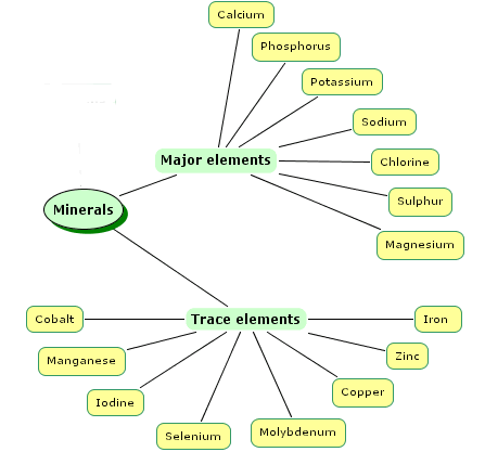
Vitamins
Rather like minerals, vitamins are needed in very small amounts in a diet but are absolutely crucial for the healthy functioning of the body. The diseases associated with a lack of a particular vitamin in the diet are called deficiency diseases. You need to know about three vitamins – A. C and D 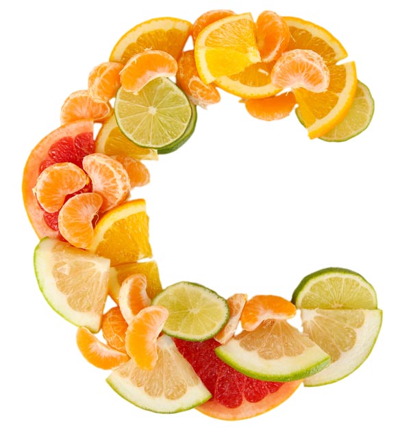 Vitamin A is a molecule called retinal found in carrots, red peppers and swede. It is needed for healthy growth and a functioning immune system. Vitamin A is also essential for normal vision since it is used to make the pigment found in rod cells in the retina. Vitamin A deficiency in the diet often causes poor vision, especially at night. Vitamin C is needed for the enzyme that produces the protein collagen in the body. It is found in all fruit especially citrus fruits. A lack of vitamin C causes the deficiency disease scurvy. Vitamin D is an unusual vitamin since it can be made in the skin using UV light. Vitamin D is needed in the small intestine to absorb mineral ions such as calcium, magnesium, etc. into the blood. A lack of vitamin D often results in a deficiency disease called rickets in which the bones malform.
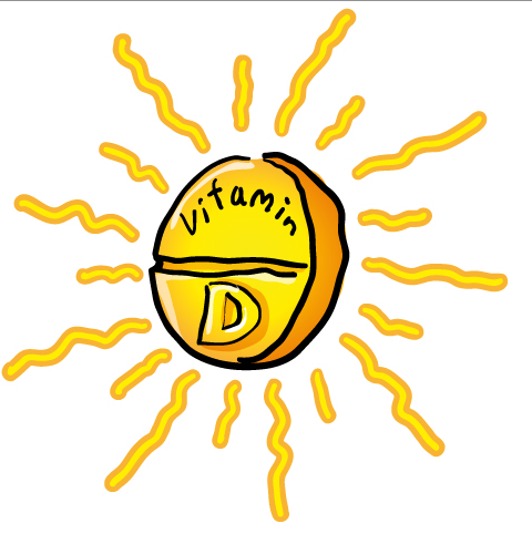
Fibre
Dietary Fibre is actually made up from the molecule cellulose. No mammal including humans possesses a cellulase enzyme and so when plant material passes through the intestines, dietary fibre is never digested. This means it passes into the large intestine where it helps prevent constipation. Foods rich in fibre included wholegrain bread, vegetables and some breakfast cereals.
Water
Water is the final component of a balanced diet. It is needed to replace water lost by sweating and in urine and acts as a solvent of course for all the metabolic reactions that happen in every cell.

Mineral ions in Plants: Grade 9 Understanding for IGCSE Biology 2.22
This posts addresses one of the commonest misconceptions you encounter as a biology teacher and it concerns a mistaken belief about the function of the roots of a plant.
The roots anchor the plant in the ground and so prevent it toppling over due to wind. But their main function is to do with the absorption of materials from the soil into the cells of the plant. The question is what exactly is taken up in the roots?
Well most people remember that water is absorbed in the roots by osmosis. The best candidates will remember the microscopic root hair cells in the root that massively increase the surface area for the uptake of water. This absorbed water is transported into the xylem tissue in the centre of the root and then moved up the plant to the leaves by transpiration pull.

Roots also absorb mineral ions from the soil by active transport. Active transport is the process where energy from respiration in the cell is used to pump material across the cell membrane against the concentration gradient. Mineral ions absorbed included nitrate ions (needed to make amino acids and proteins), magnesium ions (needed to make chlorophyll) and phosphate ions (needed to make DNA)

So where is the common misconception? This all seems sensible and fairly straightforward. Roots absorb water by osmosis and mineral ions by active transport.
Whenever root function is tested in exams, many candidates get in a pickle as they confuse mineral ions (nitrate, phosphate, magnesium, potassium) with food molecules. Plants do NOT absorb food molecules through their roots. There are very few food molecules such as glucose, amino acids, and lipids in soil. If there were, more animals would eat soil as a source of nutrition…… Plants do not need to absorb food molecules of course: the big idea you learn is that plants can make their own food molecules in the leaves in the process of photosynthesis.
So in your exam, if you ever find yourself writing anything that suggests that plants take in food through their roots, stop, take a deep breath, cross it all out and count yourself lucky you have prevented yourself from one horror answer at least!

Cell Structure: Grade 9 Understanding for iGCSE Biology 2.2 2.3 2.4
All living organisms are made from cells. Indeed the cellular nature of life is one of the universal features shared by all life on earth. Some organisms are made from just one cell (unicellular organisms) while at some point around 1 billion years ago, cells starting clumping together and specialising to form multicellular organisms such as animals and plants.
What do all cells have in common?
All cells are surrounded by a cell membrane. The cell membrane is made from a mixture of proteins and a type of lipid called a phospholipid. The cell membrane serves many functions but perhaps the most significant is acting as a partially permeable barrier that can control which molecules can enter and leave the cell.
Inside the cell membrane there is a watery solution of chemicals called the cytoplasm. The cytoplasm is the site of many metabolic reactions in the cell because many enzymes are dissolved in the cytoplasm. The cytoplasm also contains many tiny nano machines for assembling proteins called ribosomes.
And that is about it for things all cells have in common. Prokaryote cells (bacteria) have a very different cell structure with no organelles but in this section you need to understand the simplified structure of two eukaryote cells: a typical animal (on the left below) and a typical plant cell (on the right).

Both animal and plant cells have a nucleus. This is the largest organelle and contains the DNA which is the genetic material. The DNA is found in long thread-like structures called chromosomes. The nucleus controls the division of the cell and also the various functions of the cell by regulating which proteins get made.

Animal and Plant cells both contain mitochondria which are the organelles associated with aerobic respiration. Mitochondria are recognisable in the cytoplasm of the cell as sausage-shaped organelles with a folded inner membrane (see diagram above).

Structures found only in Plant cells
1) All plant cells have a thick rigid cell wall made of the carbohydrate cellulose. The cell wall allows plant cells to become turgid since when the cell takes in water by osmosis, the rigid cell wall prevents the cell from bursting. The cell wall also acts as a transport pathway across plant tissues and can provide a barrier to some pathogens.
2) All plant cells have a large permanent central sap vacuole. This organelle is bounded by a membrane called the tonoplast and in many plant cells takes up the majority of the volume of the cell.

The sap vacuole provides a compartment in the cell into which excretory molecules can be moved to stop them poisoning the cytoplasm. It also plays a role in the water balance of plant cells since because of all the solute dissolved in it, the cell sap has a low water potential. This helps draw in water by osmosis from the cytoplasm and hence from outside the cell across the cell membrane.
3) Many but not all plant cells contain chloroplasts. These are organelles associated with the process of photosynthesis. Chloroplasts can be recognised in a light microscope image as small, green structures in the cell. The green pigment comes from the chlorophyll molecules that trap energy from sunlight. In an electron micrograph, chloroplasts are distinguished due to their stacks of membrane discs called grana.


Differences between plant and animal cells


Levels of Organisation: Grade 9 Understanding for IGCSE Biology 2.1
The Easter holiday is the most important time of year for this iGCSE Biology blog. With exams in early May, the next few weeks should be the time when students are working at their maximal rate. I intend to add one post a day such that by the middle of April, the entire EdExcel iGCSE Biology specification has been covered on this blog. This should then make it a useful resource for all GCSE Biology students to help them with their revision.
Today I will put up two posts that will look at two of the simplest topics in the specification: the first one will be Levels of Organisation and then Cell Structure (2.1, 2.2, 2.3 and 2.4)
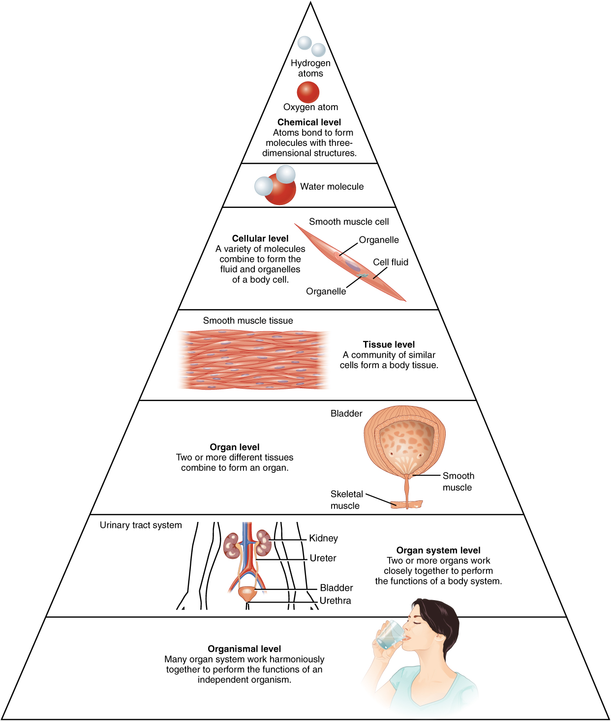
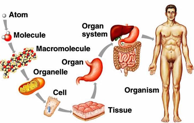
Living things (or organisms to be precise) are complex entities. Even the simplest organism will be made up of millions of different molecules arranged in an organised and complex way. Human beings are organisms made up of about 10 trillion cells of roughly 210 different cell types all put together in a organised and systematic way. It makes it much easier to study such complexity if we have a system to break the complexity down into constituent parts. This is what scientists mean by levels of organisation.
So, starting with the smallest things that might be of interest to a biologist……
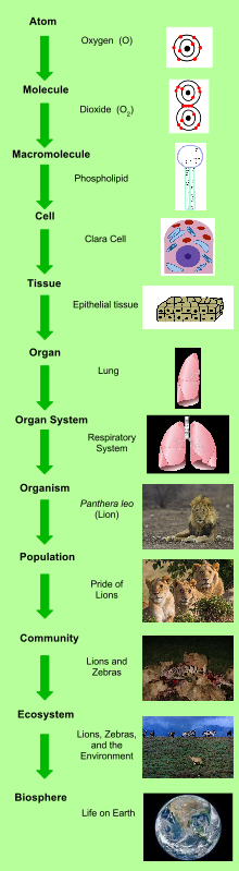 All matter on earth including the matter of living things is made of atoms (e.g. a carbon atom, an oxygen atom etc.). Atoms can combine together in a variety of ways to form molecules (a water molecule H2O, a carbon dioxide molecule CO2,) How atoms combine to form molecules is chemistry, and the levels of organisation smaller than an atom forms part of physics, so we won’t worry too much about them….
But molecules in an organism are interesting and worth studying – you learn about carbohydrates, lipids, proteins, DNA in your iGCSE course. These molecules can be grouped together to form structures inside cells called organelles. If you are being really precise with your terminology, an organelle is a membrane-bound compartment inside a eukaryotic cell (remember bacterial cells have no organelles at all). Examples of organelles are structures like the nucleus, chloroplasts, mitochondria and so on.
Cells are structures enclosed by a cell membrane that contain many different organelles. You have probably looked at a human cheek cell using a light microscope at some point in the past. In multicellular organisms, cells of the same type are often attached together to form a Tissue. A tissue is a group of similar cells often attached to each other that carry out the same function in an organism. (It is also a small disposable piece of rectangular fabric used for blowing your nose but that is something quite different….) Tissues are grouped together to form larger structures called Organs. For example, the lungs are an organ made up of a particular arrangement of epithelial tissues together with some blood and connective tissues. Organs can be grouped into Organ Systems based on their function such as the Digestive System (oesophagus, tongue, stomach, pancreas, liver, intestines etc.) An Organism such as you or I is made up of many organ systems (nervous system, cardiovascular system, digestive system, excretory system and so on….. You get the idea I’m sure!)
You can study levels of organisation bigger than the organism. This branch of biology is called Ecology – and indeed you should know the meaning of the terms population, community and ecosystem – but perhaps that is for another post……