Tagged: protease

Digestion of Proteins: Grade 9 Understanding for IGCSE Biology 2.29
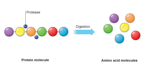
Proteins are large insoluble molecules made up of many hundreds of amino acids joined together in a long chain. So in order to obtain these molecules from our diet, the large protein must be digested (broken down) into the smaller amino acid subunits. Amino acids can be absorbed into the blood stream in the ileum, part of the small intestine.
The family of enzymes that can catalyst the digestion of proteins are called proteases.
Protein digestion happens in a two-stage process. In the first stage the large protein molecules are broken down into smaller proteins (often called polypeptides) by a protease enzyme. Pepsin is one such protease and acts in the stomach.
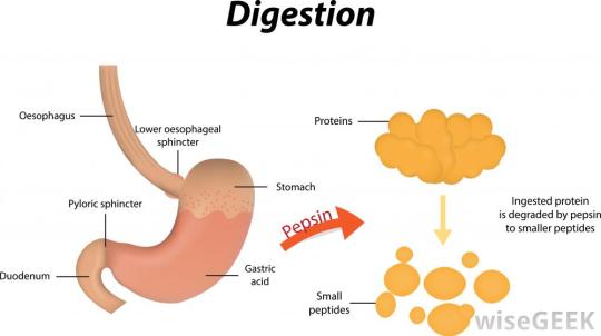
Remember that the food in the stomach is mixed with hydrochloric acid. This results in a very acidic liquid in the stomach (chyme). Pepsin works in the stomach and so rather unusually for a digestive enzyme, it has an optimum pH of pH 1.5 – pH2.
The second protease enzyme that you should know about is trypsin. Trypsin is made in the pancreas and so enters the duodenum soon after the stomach contents pass the pyloric sphincter (see diagram above). The acidic chyme that enters the duodenum is rapidly neutralised by hydrogencarbonate ions (an alkali) secreted in the bile and in pancreatic juice. Trypsin has an optimum pH of around pH 7.5.
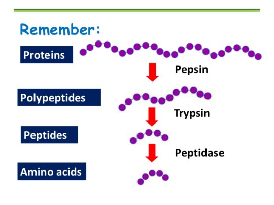
As shown in the diagram above, there is a final stage to protein digestion. The actions of pepsin in the stomach and trypsin the duodenum result in small protein fragments called peptides. Many peptides are still too large to be absorbed into the blood in the ileum and so need digesting further into their constituent amino acids. Peptidase enzymes are embedded in the epithelial cell membranes in the small intestine and this final reaction completes the digestion of proteins.
Amino acids are absorbed by active transport into the blood capillaries in the villi in the small intestine.

Small Intestine: Grade 9 Understanding for IGCSE Biology 2.32
The first part of the small intestine, called the duodenum is principally involved in digestion. Large insoluble food molecules such as proteins, lipids and starch are chemically broken down into smaller molecules in reactions catalysed by digestive enzymes.
This post will look at the longer regions of the small intestine, the jejunum and ileum. There are some digestive reactions that happen here but the main function of these parts of the intestine is the absorption of the smaller products of digestion into the body.

You should understand already which molecules are produced as products of digestion: glucose from the digestion of carbohydrates, amino acids from the breakdown of proteins and fatty acids and glycerol from the digestion of triglyceride lipids. (see my post on digestion) These then are the molecules that diffuse from the intestine into the body in the small intestine.
How is the structure of the small intestine adapted for absorption?
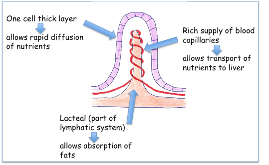
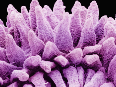
The main idea here is that the lining of these parts of the small intestine has a very large surface area. The intestine is long, the wall is ridged and the lining (called the epithelium) has many thousands of tiny projections called villi. Each villus is 1-3mm long but the effect is to increase the surface area for absorption by many hundreds of times.
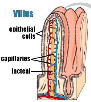
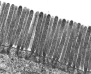
These epithelial cells have a cell membrane which is folded into many thousands of tiny structures called microvilli. Microvilli (or a brush border) can only be seem with an electron microscope and act to increase the surface area still further.
The cells that line the villus are called epithelial cells and are found in a layer that is just one cell thick. This reduces the distance the products of digestion have to move across to be absorbed.
Each villus contains a dense network of blood capillaries. This means that glucose and amino acids can easily diffuse into the blood and then be taken away from the small intestine to the liver in the hepatic portal vein. There is also a blind-ended single tube called a lacteal in each villus. This tube forms part of the lymphatic system and is used to transport fatty acids and glycerol away from the small intestine.