
Category: IGCSE Biology posts
Mutation – Grade 9 Understanding for IGCSE Biology 3.34 3.37B
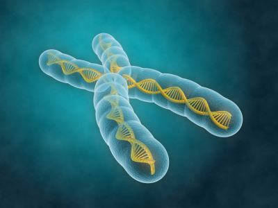
Mutations are changes in the DNA content of a cell. There are various ways the DNA of a cell could change and so mutations tend to be grouped into two main categories: chromosomal mutations and gene mutations.
Chromosomal mutation
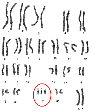
This is a change in the number or length/arrangement of the chromosomes in the nucleus. For example, people with Down’s syndrome have an extra copy of chromosome 21 giving them three chromosome 21s as opposed to the normal two.
(How many chromosomes in total will a person with Down’s syndrome have in each cell?)
Chromosomal mutations are often found in tumour cells and so play a critical role in the development of various cancers.
Sometimes the number of chromosomes in a cell stays the same, but sections are deleted, duplicated or break off from one chromosome to attach elsewhere. If this happens, this too would be classed as a chromosomal mutation.


Gene mutation
Gene mutations happen to change the sequence of base pairs that make up a single gene. As you all know, the sequence of base pairs in a gene is a code that tells the cell the sequence of amino acids to be joined together to make a protein. A gene then is the sequence of DNA that codes for a single protein. If you alter the sequence of base pairs in the DNA by adding extra ones in, or deleting some or inverting them, this will alter the protein produced.
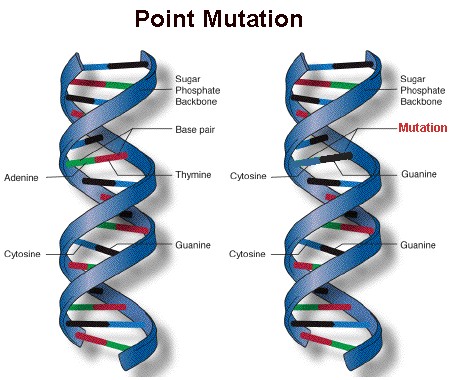
A point mutation is a change to just one base within the gene – it occurs at a single point on the DNA molecule.
Mutations can happen at any time and occur randomly whenever the DNA is replicated. But there are certain things that can increase the rate of mutation and so make harmful mutations more likely. A mutagen is an agent that increases the chance of a mutation occurring.
a) Radiation can act as a mutagen
Some parts of the electromagnetic spectrum can cause mutations when they hit DNA molecules or chromosomes. This is called ionising radiation and includes gamma rays, X rays and ultraviolet. You probably know that the dentist goes out of the room whenever they take an X ray to protect themselves from repeated exposure to X rays and you all certainly know of the link between UV exposure and incidence of skin cancer.
b) There are chemical mutagens as well
Some chemicals can make the rate of mutation increase. These are called chemical mutagens and a good example is the tar in tobacco smoke. Tar can cause cancers to form wherever the cigarette smoke comes into contact with cells and this is because tar is a mutagen. It makes mutations in the DNA much more likely and mutations are needed to turn a healthy cell into a cancer cell.
Role of the Amnion – Grade 9 Understanding for IGCSE Biology 3.12
Once the embryo has reached the uterus 7 days after fertilisation, it can implant into the thickened, sticky and blood-rich endometrium. The implanted embryo grows into the uterine lining and starts to surround itself with a collection of membranes. Some of these membranes develop into structures in the placenta, but one the amnion has a different function altogether. The amnion produces a fluid called amniotic fluid that cushions the developing embryo and foetus right through pregnancy and to birth.

The main advantage of having the developing embryo in a sac of amniotic fluid is that it protects the embryo by cushioning against blows to the abdomen. It is also essential for allowing the foetus to move around inside the uterus thus allowing development of the muscular system. The amniotic fluid enters the babies lungs and can promote normal development there. The foetus will swallow amniotic fluid into its stomach and will produce urine into the amniotic fluid as well. Disgusting I know, but that’s babies for you……..
Amniotic fluid contains stem cells. In the future it may be possible to harvest these pluripotent cells and use them to create adult tissues for medical uses.
Role of the Placenta – Grade 9 Understanding for IGCSE Biology 3.11
There are two syllabus points in bold (only tested in paper 2) that refer to embryonic and foetal development. The first asks you to understand the role of the placenta in supplying the developing foetus with nutrients and oxygen and the second concerns the role of amniotic fluid in protecting the developing embryo.
Placenta
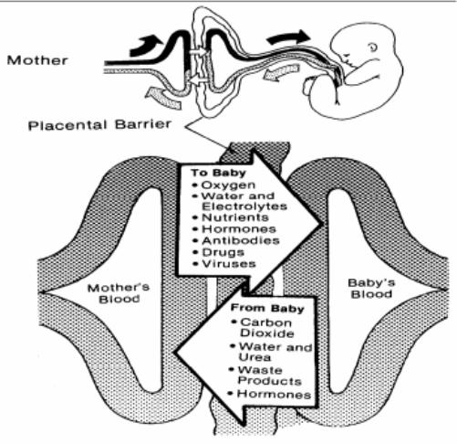
The placenta is in many ways a remarkable organ. It contains a mixture of maternal cells from the uterine lining and embryonic cells, but these cells from two genetically different individuals are capable of sticking together to form the placenta. The placenta is only present in the uterus once an embryo has successfully implanted a week or so after fertilisation has happened in the Fallopian Tubes. The placenta is linked to the foetus via the umbilical cord, a structure that contains an umbilical artery and vein carrying foetal blood to and from the placenta.

There is a key idea here that is very important. There is no mixing of maternal and foetal blood in the placenta. This would be disastrous for both mother and baby for a whole variety of reasons. The maternal blood is at a much higher pressure than the foetal blood and if the foetus were connected to the maternal circulatory system directly, its blood vessels would burst. The foetus and mother can have different blood groups of course and you may now that some blood groups are incompatible and can trigger clotting. So it is essential that there is never any mixing of blood. But what happens in the placenta is that mother’s blood empties into spaces in the placenta and babies’ blood is carried by the umbilical artery into capillaries that are found in finger-like projections called villi. This means there is a large surface area and a thin barrier between the two bloods and so exchange of materials by diffusion is possible.
The main function of the placenta then is to allow the exchange of materials between the foetal and maternal circulations. The developing foetus inside its mother’s uterus has no direct access to oxygen nor food molecules of course yet both are needed to allow healthy development. The foetus also needs a mechanism to get rid of the waste molecule, carbon dioxide that is being produced in all its cells all the time. Until the kidneys mature fully the foetus also has to get rid of urea, another excretory molecule that could build up to toxic concentrations unless removed from the growing foetus.
A few interesting points:
You will see that antibodies are small enough to cross the placenta. This gives the baby a passive immunity that can protect it for a short time from any pathogens it encounters.
Drugs such as alcohol and nicotine can cross the placenta. This is why it is so vital that pregnant mothers do not smoke and drink to ensure that the foetus’ development is not affected by these drugs.
Germination – Grade 9 Understanding for Biology GCSE 3.5 3.6
In the topic of sexual reproduction in plants, the final stage is often overlooked. I think it is helpful for students to think of this topic in several distinct stages.
- Flower Structure (hermaphrodite nature of most plants)
- Pollination (self v cross pollination; wind v insect pollinated flowers)
- Fertilisation (how does the pollen tube reach the egg cell to fertilise it?)
- Seed and Fruit formation (what forms what after fertilisation)
- Seed Dispersal (by animals, by wind, by water, by explosive means)
- Germination
Once the seed has been dispersed there then follows a period of dormancy when nothing happens. In latitudes such as the UK this often is there to delay germination until the following spring when growing conditions become more favourable. The process of taking an inert seed and growing a new plant from it is called germination.
You don’t need to worry too much about the details of germination but there are a few vital parts of the process that GCSE candidates need to appreciate for A* marks. Firstly you should know the structure of a typical seed.
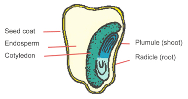
The seed coat or testa surrounds the seed and provides a tough waterproof container. Inside there are the embryonic plant (composed of a plumule and radicle), one or two seed leaves called cotyledons and a storage tissue called endosperm.
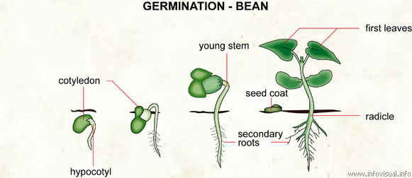
Germination starts when the seed starts to take up water by osmosis. There is an opening in the testa called the micropyle that allows water to move into the seed causing it to swell and thus rupture the seed coat to allow the embryo plant to emerge.

Water entering the seed also activates the embryo plant such that it starts to release digestive enzymes such as amylase. Amylase catalyses the digestion of starch into a simple sugar maltose. The endosperm and cotyledons contain energy stores in the form of starch, lipids and proteins and as these get broken down by the various enzymes, they provide the energy for the early growth of the seedling. The radicle emerges first and grows downwards (positive geotropism) and then the plumule or shoot grows upwards towards light (positive phototropism). Remember that throughout the early stages of this growth the energy required comes from stored food molecules in the seed. If you measure the mass of the plant during this phase, it would be decreasing. Only when the first leaves emerge above ground and the plant can start the process of photosynthesis will the overall mass start to increase.
Understanding the Eye to Grade 9 at GCSE Biology (part 2) 2.91 2.92
In the first blog post on this series, I described the pupil reflex in the eye. If you remember this involved the circular and radial muscles in the iris contracting and relaxing in an antagonistic fashion to alter the size of the pupil. You should understand why the pupil size needs to altered and what state the two sets of muscles are in varying light intensities.
But there is a second reflex in the eye totally separate from the pupil reflex and it is to do with focusing. This reflex is sometimes called accommodation but as this is a word I can’t spell, I prefer to call it focusing…. The retina at the back of the eye contains the photoreceptors. There are two types of photoreceptor in the retina (rods and cones) and these are individual cells that can detect the light and then send a nerve impulse in the optic nerve that goes to the brain.
Focusing in the Eye (this is quite complicated and needs careful, slow reading)
When the eye views objects from differing distances away, the degree the light has to be bent to produce a focused image will vary. Light coming from near objects will be diverging (the rays will be moving away from each other) and so to focus the light onto the retina, a large amount of bending (or better still refraction) will be needed. Light rays coming from far away objects are almost parallel when they hit the eye so the degree of refraction required is much less.

How can varying degrees of refraction be achieved in the eye?
Well as the diagram above shows, this is brought about by changing the shape of the lens. A short fat lens will refract (or bend) the light more than a long thin one. (If you want an explanation for why this is, you need to ask a Physics teacher – it is to do with the angle of curvature of the lens and the refractive indices of the liquids in the eye compared to the lens…….)
The lens in its default state is short and fat. This means that with no tension pulling it out of shape it will adopt the short fat shape suitable for viewing near objects.
How can the lens be pulled out of its default short fat shape?
Now this is the bit where people get confused. Read this section really carefully, check with your own notes and revision notes and make sure you have got this all the right way round! Here goes…..
There is a ring of muscle that surrounds the lens in the eye called the ciliary muscle. (Please make sure you don’t confuse this with the circular muscles in the iris) The ciliary muscle doesn’t attach to the lens directly but is attached to the lens via some strong and inelastic ligaments called the suspensory ligaments. Tension in the suspensory ligaments can pull the lens from its default short, fat shape into the long this shape needed to view far away objects.
When the ciliary muscle contracts, it shortens. This effectively moves it closer to the lens and so any tension in the suspensory ligaments is released as the ligaments go slack. Slack ligaments mean the lens adopts its short fat shape.
When the ciliary muscle relaxes, this changes its position to increase the tension in the suspensory ligaments. Taut suspensory ligaments (caused by the relaxed muscle) will pull the lens into a long thin shape.
You can easily see why people get confused here: a contracted ciliary muscle leads to slack suspensory ligaments and vice versa.

One way you can improve your understanding is to be really precise with your use of language. The ciliary muscle is a muscle (no honestly it is) and as you know, muscles can either contract or relax. Suspensory ligaments cannot contract or relax but their tension can be altered from taut (loads of tension) to slack.
So to summarise this complex sequence of events:
Looking at a far object
- Lens needs to be long and thin
- as light rays are almost parallel as they hit the eye
- and so require little bending.
- To pull the lens long and thin requires
- suspensory ligaments to be taut
- and this is achieved by the ciliary muscle relaxing.
Looking at a near object
- Lens needs to be short and fat
- as light rays are diverging as they hit the eye
- and so require a lot of bending.
- The lens will adopt a short, fat shape with
- no tension in the suspensory ligaments (the ligaments are slack)
- and this is achieved by contracting the ciliary muscle.
You can easily check your understanding here because it is much more tiring on the eye to look at a near object. If you sit on a sunny beach after all your GCSEs are finished, staring out to sea in a contemplative manner wondering how you managed to work so hard through the revision period, you could continue like this for hours. But if you try staring at your finger a few centimetres from your face for even a few seconds, your eye starts to tire. In the former scenario the ciliary muscle is relaxed and so not expending any energy but in the latter, the ciliary muscle is contracted, using energy from respiration and so can get tired.
Please comment me on this blog post with any questions – I will do my best to respond to anyone who gets in touch.
Good luck and keep working hard!
Understanding the functioning of the Eye to Grade 9 for Biology IGCSE (part 1) 2.91, 2.92
There are two reflex responses in the eye that you need to fully understand for A* levels at iGCSE. It is really easy to get them confused but I am going to put on consecutive blog posts so you can see the similarities and differences easily.
The first is a reflex called the “Pupil Reflex” which is to ensure an appropriate amount of light enters the eye in both bright and dim light. The only structure in the eye involved in the Pupil Reflex is the Iris. The second reflex explained in part 2 is the “Focusing Reflex” (or sometimes Accommodation) which makes sure that light entering the eye from objects at different distances away is focused correctly onto the retina. The structures involving in Focusing are the Lens, Ciliary Muscle and Suspensory Ligaments.
The Pupil Reflex
1) Why do we need a pupil reflex?
The eye has evolved a mechanism to ensure that the amount of light entering the eye can be adjusted. In bright light you need to limit the amount of light to prevent the light damaging the light-sensitive cells in the retina (a process called “bleaching”) and this is done by making the pupil at the front of the eye small. A small pupil would be useless for vision in low light intensities as then not enough light would get to the retina and vision would be very poor. So in dim light (low light intensities) the pupil is enlarged to allow a maximal amount of light into the eye.
2) What is the Pupil?
The pupil isn’t really a structure at all as it is simply a circular hole in the iris. The iris is a coloured muscular disc at the front of the eye.
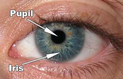
The iris has two sets of antagonistic muscles in it that can contract or relax to change the diameter of the pupil. There are radial muscles arranged like the spokes of a bicycle tyre and also circular muscles in the iris as shown in the diagram below.

3) How do the muscles in the iris bring about the pupil reflex?
Remember muscles can only contract or relax. When the radial muscles contract (shorten) they will pull the iris into a narrower shape so the pupil gets much wider. When the circular muscles contract, they will squeeze the pupil smaller so the pupil will narrow.

So you need to basic understand the state of these two sets of antagonistic muscles in both bright and dim light.
Bright light – circular muscles contracted, radial muscles relaxed, pupil small
Dim light – circular muscles relaxed, radial muscles contracted, pupil large
Gas Exchange in Plants – Grade 9 Understanding for IGCSE 2.40B, 2.41B, 2.44B, 2.45B
The topic of gas exchange in plants is often tested in exams because it can be a good discriminator between A grade and A* grade candidates. If you can master the understanding needed for these questions, important marks can be gained towards your top grade.
Firstly you must completely remove from your answers any indication that you think that plants photosynthesise in the day and respire at night. Even typing this makes me feel nauseous…. Yuk? Respiration as you all know happens in all living cells all the time and so while the first half of the statement is true (photosynthesis only happens in daytime), respiration happens at a steady rate throughout the 24 hour period.

Although the equations above make it look like these two processes are mirror images of each other, this is far from the truth.
How can gas exchange in plants be measured?
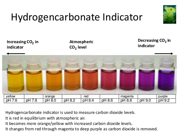
The standard set up involves using hydrogen carbonate indicator to measure changes in pH in a sealed tube. In this experiment an aquatic plant like Elodea is put into a boiling tube containing hydrogen carbonate indicator. The indicator changes colour depending on the pH as shown below:
- acidic pH: indicator goes yellow
- neutral pH: indicator is orange
- alkaline pH: indicator goes purple
a) If the tube with the plant is kept in the dark (perhaps by wrapping silver foil round the boiling tube), what colour do you think the indicator will turn? Explain why you think this.
b) If the tube with the plant is kept in bright light, what colour do you think the indicator will turn and why?
c) If a control tube is set up with no plant in at all but left for two days and no colour change is observed, what does this show?
In order to score all the marks on these kind of questions, there are two pieces of information/knowledge you need to demonstrate. You need to show the examiner that you understand that carbon dioxide is an acidic gas (it reacts with water to form carbonic acid) and so the more carbon dioxide there is in a tube, the more acidic will be the pH. As oxygen concentrations change in a solution, there will be no change to the indicator as oxygen does not alter the pH of a solution.
Secondly you need to show that you understand it is the balance between the rates of photosynthesis and respiration that alters the carbon dioxide concentration. If rate of respiration is greater than the rate of photosynthesis, there will be a net release of carbon dioxide so the pH will fall (become more acidic). If the rate of photosynthesis in the tube is greater than the rate of respiration, there will be a net uptake of carbon dioxide (more will be used in photosynthesis than is produced in respiration) and so the solution will become more alkaline.
So to answer the three questions above I would write:
a) The indicator will turn yellow in these conditions. This is because there is no light so the plant cannot photosynthesise but it continues to respire. Respiration releases carbon dioxide as a waste product so because the rate of respiration is greater than the rate of photosynthesis, there will be a net release of carbon dioxide from the plant. Carbon dioxide is an acidic gas so the pH in the solution will fall, hence the yellow colour of the solution.
b) The indicator will turn purple in these conditions. This is because the bright light means the plant photosynthesises at a fast rate. Photosynthesis uses up carbon dioxide from the water. The plant continues to respire as well and respiration releases carbon dioxide as a waste product. As the rate of photosynthesis is greater than the rate of respiration in these conditions there will be a net uptake of carbon dioxide. Carbon dioxide is an acidic gas so if more is taken from the solution than released into it, the pH in the solution will rise as it becomes more alkaline, hence the purple colour of the solution.
c) This shows that without a living plant in the tube there is nothing else that can alter the pH of the solution. It provides evidence that my explanations above about the cause of the colour change is correct.
Skin – Grade 9 Understanding for IGCSE Biology 2.93
The skin as you all know is the largest organ in the human body. It has a variety of functions including providing a water-tight barrier to minimise evaporation from the cells; it is also a habitat for billions of bacteria that live on the skin, the so-called skin flora and it contains a variety of sensory receptors that provide information about the external world to our central nervous systems. The iGCSE specification requires you to know about the role of skin in thermoregulation.
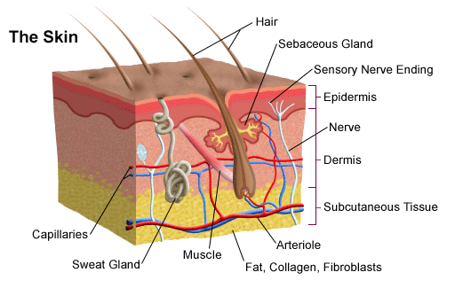
The skin is made up of an outer layer of dead cells called the epidermis that contains sensory nerve endings. Beneath this is the dermis which is made of living cells and blood vessels, sweat glands, hair follicles and other specialised sensory receptors, e.g. for touch. Underneath the dermis there is a subcutaneous tissue that in humans is packed full of adipose cells that store lipids.
The skin is involved in thermoregulation both as a receptor and more significantly as an effector.
The skin’s role as a receptor in thermoregulation
The brain receives information about temperature from two sets of thermoreceptors. There are receptors in the hypothalamus that measure the temperature of the blood passing through the brain. This provides information about core body temperature. In the skin there are two types of thermoreceptors, called hot and cold receptors, that together monitor the external temperature. Information from both these sets of receptors is used by thermoregulatory centres in the hypothalamus to regulate your body temperature.
The skin’s role as an effector in thermoregulation
The skin is the principle effector organ for thermoregulation. This is because it is found at the boundary between your cells and the external environment and so heat gain and heat loss happen through it. The skin has three ways of altering the heat gain/loss depending on nerve impulses from the CNS.
1) Sweating
Humans have sweat glands spread over almost all the surface of the skin. These glands secrete a watery liquid, sweat that contains a solution of salts and a tiny amount of urea dissolved in large volumes of water. Sweat is only produced when the body temperature is too high as the evaporation of sweat from the surface of the skin leads to a cooler skin. How does this process work?
The main idea to understand is that the sweat itself is not in any way cool. Sweat is made in sweat glands from blood plasma so if the blood is getting too hot, the sweat will be hot as well. But it takes energy to evaporate water (to turn it from the liquid to the vapour state) and this energy (called the latent heat of vapourisation) is taken as heat energy from the skin. So as sweat evaporates, it uses thermal energy from the skin to turn the water molecules in sweat into a vapour. This evaporative cooling leaves the skin cooler once the sweat has evaporated than it was at the start.
2) Hairs
Hairs on the skin play an important role in thermoregulation in many mammals but not really in our species. If the body temperature drops, the CNS causes hair erector muscles to contract and pull the hair to a more vertical position in the follicle. If an animal’s hairs stand on end, a thicker layer of air is trapped between them and so the body is better insulated against heat loss. Humans are relatively hairless and the only thing that really happens in us when the hair erector muscles contract is that we get “goose bumps”.
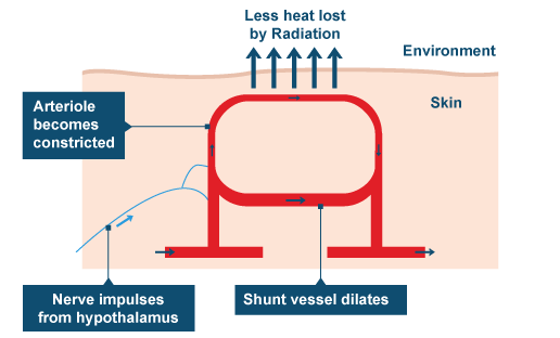
3) Shifting patterns of Blood flow in the skin
This is the main effector mechanism in human thermoregulation but it is also the one that tends to catch exam candidates out. Please make sure you understand this process fully and can explain this section of work very well indeed. If the body is getting too cold, the pattern of blood flow switches in the skin so less blood flows in the capillary beds near the surface of the skin and more blood is retained deeper in the skin structure. This is achieved by narrowing the arterioles that supply the capillary beds near the surface (arterioles and arteries have plenty of muscle in their walls that can contract to narrow the lumen of the blood vessel) This narrowing of arterioles is called vasoconstriction.
The converse happens when the body is getting too warm. The muscle in the walls of these arterioles now relaxes to widen the lumen, thus allowing more blood to flow in capillary beds near the surface. This vasodilation allows more heat to be lost from the blood by conduction, convection and radiation and so the blood leaving the skin has lost more heat to the external environment.
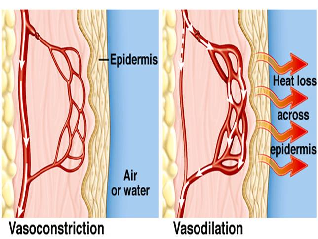
You will notice that at no point in these explanations of vasoconstriction and vasodilation do I mention capillaries in the skin moving deeper or nearer the surface. For some reason every year, GCSE candidates think that the reason you look redder when you are hot is because capillaries in the skin move nearer the surface. This cannot be true – blood vessels have a fixed position in the body for a start – but now you should understand that you look redder when you are hot because the capillaries that happen to be near the surface are having a greater volume of blood per minute flowing through them because of vasodilation. If you find yourself in the exam writing about capillaries moving in response to a change in temperature, please stop writing, take a deep breath, count to ten and then cross it all out and start again!
How is energy lost between one trophic level and the next? Grade 9 Understanding for IGCSE Biology 4.8 4.9
 The diagram above shows how energy moves up the food chain through feeding. Remember that if you are asked what the arrows represent in a food chain, there is only one possible correct response. “Arrows in a food chain show the flow of energy from one trophic level to the next”
The big idea here is that not all the energy in one trophic level can ever pass to the next. The specification suggests that only 10% of the energy is transferred from one level to the next (but in fact the percentage varies between 0.1% to around 15%)
 kCal is a unit of energy and the pyramid shows that only 10% of the energy in one level is found in the next.
So there is a big question here – where does all the other 90% of the energy in one level end up?
There are a whole load of different ways energy is lost. Consider the transfer of energy between mice and owls.
- The mice use up energy in the process of respiration. The glucose molecules that mice oxidise to provide the energy to move around are not available to an owl if the mouse is eaten.
- Not all mice are eaten by owls or other predators. Many die of disease, starvation and exposure and a few might even live long enough to die peacefully in their sleep. All these “dead mice” will have energy in their bodies that cannot pass up a food chain but instead passes to decomposers.
- Even the mice that are eaten by owls are not eaten in their entirety. The owl might only eat the energy-rich parts of a mouse and regurgitate out the bones and fur. So some energy is lost as not all the mouse is eaten and digested by the owl.
- There will be parts of the mouse that even when swallowed and digested are not accessible. Owl faeces will contain some molecules from the mice eaten that contain energy. This energy is perhaps found in molecules that the owl digestive system cannot digest. The energy present in the owl faeces is lost to the food chain and like the example above will pass to decomposers.
This energy adds up to around 90% of the energy in any trophic level. Ultimately though where does it all go? All the energy in all the organisms in an ecosystem has the same fate: it ends up as heat that is dissipated into the system. Energy can only enter an ecosystem in one way (as sunlight trapped in the process of photosynthesis in producers) and in the end, it all ends up leaving the system as heat energy. This heat energy is a waste product of respiration.
Xylem transport – Grade 9 Understanding for IGCSE Biology 2.54, 2.55B, 2.56B
The topic of plant transport can appear quite complicated but you will see from your past paper booklets that the questions examiners tend to set on it are much more straightforward.
The key piece of understanding is to realise that there are two transport systems in plants, learn their names and what they transport.
- Xylem vessels move water and mineral ions from the roots to the leaves.
- Phloem sieve tubes move sugars, notably sucrose, and amino acids around the plant. Both of these molecules are made in photosynthesis in the leaves and so can be transported from the leaves to the areas in the plant where they are needed.
Water is needed for photosynthesis of course in the leaves (remember that rain water cannot enter leaves directly because of the waxy cuticle on the surface of the leaf). All the water that is used in photosynthesis is absorbed in the roots from the soil and moved up the plant in the xylem vessels. Minerals such as nitrate, phosphate and magnesium ions are also required in the leaves for making amino acids, DNA and chlorophyll respectively. These minerals are moved up the plant along with the water in the xylem.
How does water enter the roots from the soil?
Water molecules can only enter root hair cells (and indeed can only cross any cell membrane) by one mechanism and that is OSMOSIS. If you understand the mechanism of osmosis that is great but don’t worry too much about it at this stage. You need to know that osmosis is a net movement of water from a dilute solution to a more concentrated solution across a partially permeable membrane.
How do mineral ions enter the roots from the soil?
Minerals are pumped into the root hair cells from the soil using ACTIVE TRANSPORT. This a process that uses energy from respiration in the cell to move ions against their concentration gradient (so from a lower concentration in the soil to a higher concentration inside the cell cytoplasm.)
What do we know about xylem vessels?
The cells that water and minerals are transported in are called xylem vessels. They have some interesting specialisations for this function. They are dead cells that are empty with no cytoplasm or nucleus. The end walls of these cells break down to provide a continuous unbroken column of water all the way up the plant. The cell walls of xylem vessels are thick and strengthened and waterproofed with a chemical called lignin.
What causes the water to move up the xylem?
Clearly it will take energy from somewhere to move water against gravity all the way up a plant from the roots to the leaves. The key question here is what provides the energy for this movement? There is no pumping of water up the plant and indeed the plant spends no energy at all on water movement. The answer is that it is the heat energy from the sun that evaporates water in the leaves that provides the energy for water movement. When you combine this with the fact that water molecules are “sticky” – they are attracted to their neighbours by a type of weak bond called a hydrogen bond – you can see that the water evaporating into the air spaces in the leaf can pull water molecules up the continuous column of water found in the xylem. The proper adjective for this stickiness is cohesive and you should know the name for the evaporation of water in the leaves (Transpiration)

